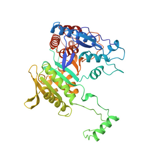
Access to phosphorylation in isocitrate dehydrogenase may occur by domain shifting.
Finer-Moore, J., Tsutakawa, S.E., Cherbavaz, D.R., LaPorte, D.C., Koshland Jr., D.E., Stroud, R.M.(1997) Biochemistry 36: 13890-13896
- PubMed: 9374867
- DOI: https://doi.org/10.1021/bi9711691
- Primary Citation of Related Structures:
1SJS - PubMed Abstract:
To clarify further the mechanism of regulation by phosphorylation of isocitrate dehydrogenase, cocrystallization of isocitrate dehydrogenase and isocitrate dehydrogenase kinase/phosphatase in the presence of an ATP analog was attempted. Although cocrystallization was unsuccessful, a new crystal form of isocitrate dehydrogenase was obtained which provides insight into the phosphorylation mechanism. The new, orthorhombic crystal form of isocitrate dehydrogenase is related to the previously reported tetragonal form largely by an approximately 16 degrees shift of a large domain relative to the small domain and clasp region within each subunit of the dimeric enzyme. The NADP+ cofactor binding surface is significantly disrupted by the shift to the open conformation. The solvent-accessible surface area and surface-enclosed volume increase by 2% relative to the dimeric tetragonal form. Most of the increase results from expansion of the active site cleft such that the distance across its opening increases from approximately 5 to 13 A, significantly increasing accessibility to Ser-113. The conformation of isocitrate dehydrogenase in the orthorhombic crystal form more closely resembles that of the crystal structure of the homologous enzyme 3-isopropylmalate dehydrogenase than does the tetragonal isocitrate dehydrogenase conformation. Since the crystal lattice forces are fairly weak, it appears that isocitrate dehydrogenase is a flexible molecule that can easily undergo domain shifts and possibly other induced fit conformational changes, to accommodate binding to isocitrate dehydrogenase kinase/phosphatase.
Organizational Affiliation:
Department of Biochemistry and Biophysics, University of California, San Francisco 94143-0448, USA.