1SBB

T-CELL RECEPTOR BETA CHAIN COMPLEXED WITH SUPERANTIGEN SEB


Experimental Data Snapshot

  • Method: X-RAY DIFFRACTION
  • Resolution: 2.40 Å
  • R-Value Free: 0.309 
  • R-Value Work: 0.228 

wwPDB Validation   3D Report Full Report


This is version 1.4 of the entry. See complete history


Literature

Three-dimensional structure of the complex between a T cell receptor beta chain and the superantigen staphylococcal enterotoxin B.

Li, H.Llera, A.Tsuchiya, D.Leder, L.Ysern, X.Schlievert, P.M.Karjalainen, K.Mariuzza, R.A.

(1998) Immunity 9: 807-816

  • DOI: https://doi.org/10.1016/s1074-7613(00)80646-9
  • Primary Citation of Related Structures:  
    1SBB

  • PubMed Abstract: 

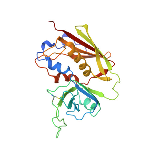
    Superantigens (SAGs) are a class of immunostimulatory proteins of bacterial or viral origin that activate T cells by binding to the V beta domain of the T cell antigen receptor (TCR). The three-dimensional structure of the complex between a TCR beta chain (mouse V beta8.2) and the SAG staphylococcal enterotoxin B (SEB) at 2.4 A resolution reveals why SEB recognizes only certain V beta families, as well as why only certain SAGs bind mouse V beta8.2. Models of the TCR-SEB-peptide/MHC class II complex indicate that V alpha interacts with the MHC beta chain in the TCR-SAG-MHC complex. The extent of the interaction is variable and is largely determined by the geometry of V alpha/V beta domain association. This variability can account for the preferential expression of certain V alpha regions among T cells reactive with SEB.


  • Organizational Affiliation

    Center for Advanced Research in Biotechnology, University of Maryland Biotechnology Institute, Rockville 20850, USA.


Macromolecules
Find similar proteins by:  (by identity cutoff)  |  3D Structure
Entity ID: 1
MoleculeChains Sequence LengthOrganismDetailsImage
PROTEIN (14.3.D T CELL ANTIGEN RECEPTOR)
A, C
238Mus musculusMutation(s): 3 
UniProt
Find proteins for P01852 (Mus musculus)
Explore P01852 
Go to UniProtKB:  P01852
Entity Groups  
Sequence Clusters30% Identity50% Identity70% Identity90% Identity95% Identity100% Identity
UniProt GroupP01852
Sequence Annotations
Expand
  • Reference Sequence
Find similar proteins by:  (by identity cutoff)  |  3D Structure
Entity ID: 2
MoleculeChains Sequence LengthOrganismDetailsImage
PROTEIN (STAPHYLOCOCCAL ENTEROTOXIN B)
B, D
239Staphylococcus aureusMutation(s): 0 
UniProt
Find proteins for P01552 (Staphylococcus aureus)
Explore P01552 
Go to UniProtKB:  P01552
Entity Groups  
Sequence Clusters30% Identity50% Identity70% Identity90% Identity95% Identity100% Identity
UniProt GroupP01552
Sequence Annotations
Expand
  • Reference Sequence
Experimental Data & Validation

Experimental Data

  • Method: X-RAY DIFFRACTION
  • Resolution: 2.40 Å
  • R-Value Free: 0.309 
  • R-Value Work: 0.228 
  • Space Group: P 1 21 1
Unit Cell:
Length ( Å )Angle ( ˚ )
a = 71.2α = 90
b = 83.65β = 93.04
c = 82.98γ = 90
Software Package:
Software NamePurpose
DENZOdata reduction
SCALEPACKdata scaling
AMoREphasing
X-PLORrefinement

Structure Validation

View Full Validation Report


Entry History 

Deposition Data

Revision History  (Full details and data files)

  • Version 1.0: 1999-03-01
    Type: Initial release
  • Version 1.1: 2008-04-26
    Changes: Version format compliance
  • Version 1.2: 2011-07-13
    Changes: Version format compliance
  • Version 1.3: 2017-10-04
    Changes: Advisory, Refinement description
  • Version 1.4: 2023-08-23
    Changes: Advisory, Data collection, Database references, Derived calculations, Refinement description