1S7Z

Structure of Ocr from Bacteriophage T7


Experimental Data Snapshot

  • Method: X-RAY DIFFRACTION
  • Resolution: 1.83 Å
  • R-Value Free: 0.239 
  • R-Value Work: 0.170 
  • R-Value Observed: 0.173 

wwPDB Validation   3D Report Full Report


This is version 1.2 of the entry. See complete history


Literature

Structure of Ocr from Bacteriophage T7, a Protein that Mimics B-Form DNA

Walkinshaw, M.D.Taylor, P.Sturrock, S.S.Atanasiu, C.Berg, T.Henderson, R.M.Edwardson, J.M.Dryden, D.T.

(2002) Mol Cell 9: 187-194

  • DOI: https://doi.org/10.1016/s1097-2765(02)00435-5
  • Primary Citation of Related Structures:  
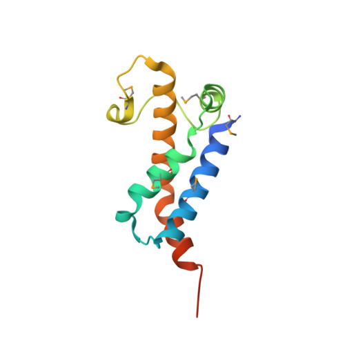
    1S7Z

  • PubMed Abstract: 

    We have solved, by X-ray crystallography to a resolution of 1.8 A, the structure of a protein capable of mimicking approximately 20 base pairs of B-form DNA. This ocr protein, encoded by gene 0.3 of bacteriophage T7, mimics the size and shape of a bent DNA molecule and the arrangement of negative charges along the phosphate backbone of B-form DNA. We also demonstrate that ocr is an efficient inhibitor in vivo of all known families of the complex type I DNA restriction enzymes. Using atomic force microscopy, we have also observed that type I enzymes induce a bend in DNA of similar magnitude to the bend in the ocr molecule. This first structure of an antirestriction protein demonstrates the construction of structural mimetics of long segments of B-form DNA.


  • Organizational Affiliation

    Institute of Cell and Molecular Biology, The King's Buildings, University of Edinburgh, EH9 3JR, Edinburgh, United Kingdom. malcolm.walkinsahaw@ed.ac.uk


Macromolecules
Find similar proteins by:  (by identity cutoff)  |  3D Structure
Entity ID: 1
MoleculeChains Sequence LengthOrganismDetailsImage
Gene 0.3 protein117Escherichia phage T7Mutation(s): 6 
Gene Names: 0.3
UniProt
Find proteins for P03775 (Escherichia phage T7)
Explore P03775 
Go to UniProtKB:  P03775
Entity Groups  
Sequence Clusters30% Identity50% Identity70% Identity90% Identity95% Identity100% Identity
UniProt GroupP03775
Sequence Annotations
Expand
  • Reference Sequence
Small Molecules
Modified Residues  1 Unique
IDChains TypeFormula2D DiagramParent
MSE
Query on MSE
A
L-PEPTIDE LINKINGC5 H11 N O2 SeMET
Experimental Data & Validation

Experimental Data

  • Method: X-RAY DIFFRACTION
  • Resolution: 1.83 Å
  • R-Value Free: 0.239 
  • R-Value Work: 0.170 
  • R-Value Observed: 0.173 
  • Space Group: C 1 2 1
Unit Cell:
Length ( Å )Angle ( ˚ )
a = 78.777α = 90
b = 37.665β = 98.42
c = 35.49γ = 90
Software Package:
Software NamePurpose
REFMACrefinement
DENZOdata reduction
CCP4data scaling
SOLVEphasing

Structure Validation

View Full Validation Report


Entry History 

Deposition Data

Revision History  (Full details and data files)

  • Version 1.0: 2004-02-10
    Type: Initial release
  • Version 1.1: 2008-04-29
    Changes: Version format compliance
  • Version 1.2: 2011-07-13
    Changes: Derived calculations, Version format compliance