


Crystal Structure of Guanylate Kinase from Mycobacterium tuberculosis
Chan, S., Sawaya, M.R., Perry, L.J., Eisenberg, D.To be published.
Experimental Data Snapshot
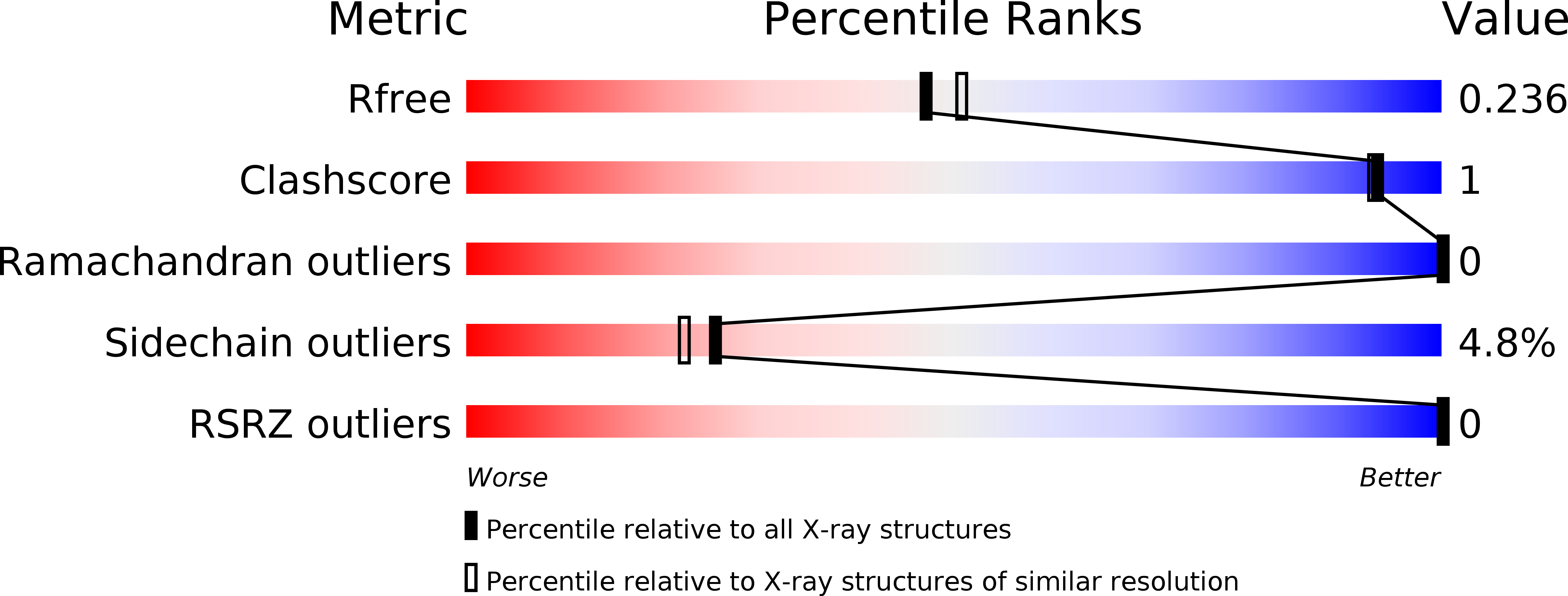
wwPDB Validation 3D Report Full Report
Entity ID: 1 | |||||
|---|---|---|---|---|---|
| Molecule | Chains | Sequence Length | Organism | Details | Image |
| Guanylate kinase | 228 | Mycobacterium tuberculosis H37Rv | Mutation(s): 0 Gene Names: GMK, RV1389, MT1434, MTCY21B4.06, MB1424 EC: 2.7.4.8 |  | |
UniProt | |||||
Find proteins for P9WKE9 (Mycobacterium tuberculosis (strain ATCC 25618 / H37Rv)) Explore P9WKE9 Go to UniProtKB: P9WKE9 | |||||
Entity Groups | |||||
| Sequence Clusters | 30% Identity50% Identity70% Identity90% Identity95% Identity100% Identity | ||||
| UniProt Group | P9WKE9 | ||||
Sequence AnnotationsExpand | |||||
| |||||
| Ligands 2 Unique | |||||
|---|---|---|---|---|---|
| ID | Chains | Name / Formula / InChI Key | 2D Diagram | 3D Interactions | |
| FMT Query on FMT | C [auth A], D [auth A], E [auth A] | FORMIC ACID C H2 O2 BDAGIHXWWSANSR-UHFFFAOYSA-N |  | ||
| CL Query on CL | B [auth A] | CHLORIDE ION Cl VEXZGXHMUGYJMC-UHFFFAOYSA-M |  | ||
| Length ( Å ) | Angle ( ˚ ) |
|---|---|
| a = 112.126 | α = 90 |
| b = 112.126 | β = 90 |
| c = 112.126 | γ = 90 |
| Software Name | Purpose |
|---|---|
| REFMAC | refinement |
| DENZO | data reduction |
| SCALEPACK | data scaling |
| MLPHARE | phasing |
RCSB PDB (citation) is hosted by
RCSB PDB is a member of the

