1RPW

Crystal Structure Of The Multidrug Binding Protein Qacr Bound To The Diamidine Hexamidine


Experimental Data Snapshot

  • Method: X-RAY DIFFRACTION
  • Resolution: 2.90 Å
  • R-Value Free: 0.280 
  • R-Value Work: 0.229 
  • R-Value Observed: 0.229 

wwPDB Validation   3D Report Full Report


Ligand Structure Quality Assessment 


This is version 1.4 of the entry. See complete history


Literature

Crystal structures of QacR-diamidine complexes reveal additional multidrug-binding modes and a novel mechanism of drug charge neutralization.

Murray, D.S.Schumacher, M.A.Brennan, R.G.

(2004) J Biol Chem 279: 14365-14371

  • DOI: https://doi.org/10.1074/jbc.M313870200
  • Primary Citation of Related Structures:  
    1RKW, 1RPW

  • PubMed Abstract: 

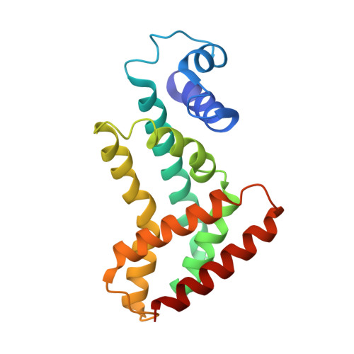
    The Staphylococcus aureus multidrug-binding protein QacR represses transcription of the plasmid-encoded membrane protein QacA, a multidrug efflux transporter. QacR is induced by multiple structurally dissimilar monovalent and bivalent cationic lipophilic compounds, many of which are effluxed from the cell by QacA via the proton motive force. The multidrug-binding pocket of QacR has been shown to be quite extensive and features several glutamates and multiple aromatic residues. To date, the structure of only one QacR-bivalent cationic drug complex (that of QacR bound to dequalinium) has been determined, and how other longer or shorter bivalent cationic compounds bind is unknown. Here we report the crystal structures of QacR bound to two cytotoxic bivalent diamidines, pentamidine and hexamidine. These compounds are structurally similar, differing by only one methylene carbon in the alkyl chain linker. However, this small difference results in very dissimilar binding modes. Similar to dequalinium, hexamidine spans the multidrug-binding pocket, and its positively charged benzamidine groups are neutralized by residues Glu-57 and Glu-120. Pentamidine binds QacR in a novel fashion whereby one of its benzamidine groups interacts with residue Glu-63, and the other is neutralized by carbonyl and side chain oxygen atoms. Thus, these structures demonstrate that a formal negative charge is not a prerequisite for binding positively charged drugs and underscore the versatility of the QacR and, likely, all multidrug-binding pockets.


  • Organizational Affiliation

    Department of Biochemistry and Molecular Biology, Oregon Health and Science University, Portland, Oregon 97239-3098, USA.


Macromolecules
Find similar proteins by:  (by identity cutoff)  |  3D Structure
Entity ID: 1
MoleculeChains Sequence LengthOrganismDetailsImage
Transcriptional regulator qacR
A, B, C, D
188Staphylococcus aureusMutation(s): 2 
Gene Names: QACRSAVP031
UniProt
Find proteins for P0A0N4 (Staphylococcus aureus)
Explore P0A0N4 
Go to UniProtKB:  P0A0N4
Entity Groups  
Sequence Clusters30% Identity50% Identity70% Identity90% Identity95% Identity100% Identity
UniProt GroupP0A0N4
Sequence Annotations
Expand
  • Reference Sequence
Small Molecules
Ligands 2 Unique
IDChains Name / Formula / InChI Key2D Diagram3D Interactions
DID
Query on DID

Download Ideal Coordinates CCD File 
K [auth B]4,4'[1,6-HEXANEDIYLBIS(OXY)]BISBENZENECARBOXIMIDAMIDE
C20 H26 N4 O2
OQLKNTOKMBVBKV-UHFFFAOYSA-N
SO4
Query on SO4

Download Ideal Coordinates CCD File 
E [auth A]
F [auth A]
G [auth B]
H [auth B]
I [auth B]
E [auth A],
F [auth A],
G [auth B],
H [auth B],
I [auth B],
J [auth B],
L [auth C],
M [auth C],
N [auth C],
O [auth C],
P [auth C],
Q [auth C],
R [auth D],
S [auth D],
T [auth D],
U [auth D],
V [auth D],
W [auth D],
X [auth D],
Y [auth D]
SULFATE ION
O4 S
QAOWNCQODCNURD-UHFFFAOYSA-L
Experimental Data & Validation

Experimental Data

  • Method: X-RAY DIFFRACTION
  • Resolution: 2.90 Å
  • R-Value Free: 0.280 
  • R-Value Work: 0.229 
  • R-Value Observed: 0.229 
  • Space Group: P 42 21 2
Unit Cell:
Length ( Å )Angle ( ˚ )
a = 171.8α = 90
b = 171.8β = 90
c = 94.5γ = 90
Software Package:
Software NamePurpose
CNSrefinement
MOSFLMdata reduction
CCP4data scaling
CNSphasing

Structure Validation

View Full Validation Report


Ligand Structure Quality Assessment 


Entry History 

Deposition Data

Revision History  (Full details and data files)

  • Version 1.0: 2004-06-01
    Type: Initial release
  • Version 1.1: 2008-04-29
    Changes: Version format compliance
  • Version 1.2: 2011-07-13
    Changes: Version format compliance
  • Version 1.3: 2021-10-27
    Changes: Database references, Derived calculations
  • Version 1.4: 2023-08-23
    Changes: Data collection, Refinement description