1RFT

Crystal structure of pyridoxal kinase complexed with AMP-PCP and pyridoxamine


Experimental Data Snapshot

  • Method: X-RAY DIFFRACTION
  • Resolution: 2.80 Å
  • R-Value Free: 0.268 
  • R-Value Work: 0.214 
  • R-Value Observed: 0.218 

wwPDB Validation   3D Report Full Report


Ligand Structure Quality Assessment 


This is version 1.3 of the entry. See complete history


Literature

Conformational changes in the reaction of pyridoxal kinase

Li, M.-H.Kwok, F.Chang, W.-R.Liu, S.-Q.Lo, S.C.L.Zhang, J.-P.Jiang, T.Liang, D.-C.

(2004) J Biol Chem 279: 17459-17465

  • DOI: https://doi.org/10.1074/jbc.M312380200
  • Primary Citation of Related Structures:  
    1RFT, 1RFU, 1RFV

  • PubMed Abstract: 

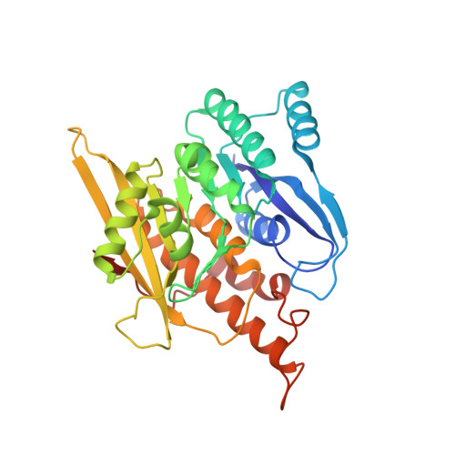
    To understand the processes involved in the catalytic mechanism of pyridoxal kinase (PLK),1 we determined the crystal structures of PLK.AMP-PCP-pyridoxamine, PLK.ADP.PLP, and PLK.ADP complexes. Comparisons of these structures have revealed that PLK exhibits different conformations during its catalytic process. After the binding of AMP-PCP (an analogue that replaced ATP) and pyridoxamine to PLK, this enzyme retains a conformation similar to that of the PLK.ATP complex. The distance between the reacting groups of the two substrates is 5.8 A apart, indicating that the position of ATP is not favorable to spontaneous transfer of its phosphate group. However, the structure of PLK.ADP.PLP complex exhibited significant changes in both the conformation of the enzyme and the location of the ligands at the active site. Therefore, it appears that after binding of both substrates, the enzyme-substrate complex requires changes in the protein structure to enable the transfer of the phosphate group from ATP to vitamin B(6). Furthermore, a conformation of the enzyme-substrate complex before the transition state of the enzymatic reaction was also hypothesized.


  • Organizational Affiliation

    National Laboratory of Biomacromolecules, Institute of Biophysics, Chinese Academy of Sciences, Beijing 100101, China.


Macromolecules
Find similar proteins by:  (by identity cutoff)  |  3D Structure
Entity ID: 1
MoleculeChains Sequence LengthOrganismDetailsImage
Pyridoxal kinase312Ovis ariesMutation(s): 0 
EC: 2.7.1.35
UniProt
Find proteins for P82197 (Ovis aries)
Explore P82197 
Go to UniProtKB:  P82197
Entity Groups  
Sequence Clusters30% Identity50% Identity70% Identity90% Identity95% Identity100% Identity
UniProt GroupP82197
Sequence Annotations
Expand
  • Reference Sequence
Experimental Data & Validation

Experimental Data

  • Method: X-RAY DIFFRACTION
  • Resolution: 2.80 Å
  • R-Value Free: 0.268 
  • R-Value Work: 0.214 
  • R-Value Observed: 0.218 
  • Space Group: P 31 2 1
Unit Cell:
Length ( Å )Angle ( ˚ )
a = 103.687α = 90
b = 103.687β = 90
c = 58.604γ = 120
Software Package:
Software NamePurpose
DENZOdata reduction
SCALEPACKdata scaling
AMoREphasing
CNSrefinement

Structure Validation

View Full Validation Report


Ligand Structure Quality Assessment 


Entry History 

Deposition Data

Revision History  (Full details and data files)

  • Version 1.0: 2004-04-27
    Type: Initial release
  • Version 1.1: 2008-04-29
    Changes: Version format compliance
  • Version 1.2: 2011-07-13
    Changes: Version format compliance
  • Version 1.3: 2023-10-25
    Changes: Data collection, Database references, Derived calculations, Refinement description