1REV

HIV-1 REVERSE TRANSCRIPTASE


Experimental Data Snapshot

  • Method: X-RAY DIFFRACTION
  • Resolution: 2.60 Å
  • R-Value Work: 0.224 
  • R-Value Observed: 0.224 

wwPDB Validation   3D Report Full Report


This is version 1.2 of the entry. See complete history


Literature

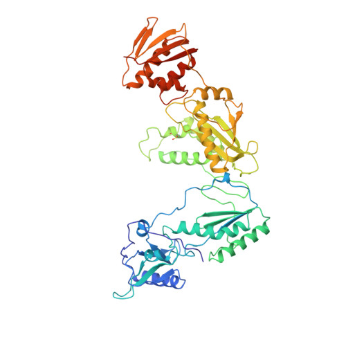
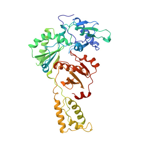
The structure of HIV-1 reverse transcriptase complexed with 9-chloro-TIBO: lessons for inhibitor design.

Ren, J.Esnouf, R.Hopkins, A.Ross, C.Jones, Y.Stammers, D.Stuart, D.

(1995) Structure 3: 915-926

  • DOI: https://doi.org/10.1016/S0969-2126(01)00226-X
  • Primary Citation of Related Structures:  
    1REV

  • PubMed Abstract: 

    HIV reverse transcriptase (RT) is a key target of anti-AIDS therapies. Structural studies of HIV-1 RT, unliganded and complexed with different non-nucleoside inhibitors (NNIs), have pointed to a common mode of binding and inactivation through distortion of the polymerase catalytic site by NNIs containing two hinged rings. The mode of binding of the TIBO family of inhibitors is of interest because these compounds do not fit the two-hinged-ring model. The structure of HIV-1 RT complexed with 9-chloro-TIBO (R82913) has been determined at 2.6 A resolution. As reported for the lower resolution analysis of another TIBO compound, this inhibitor binds at the same site as other NNIs, but our higher resolution study reveals the Cl-TIBO is distorted from the conformation seen in crystals of the inhibitor alone. This allows Cl-TIBO to mimic the binding of NNIs containing two hinged rings. Inhibitor-protein interactions are again predominantly hydrophobic and the protein conformation corresponds to that seen in complexes with other tight-binding NNIs. Although Cl-TIBO is chemically very different from other NNIs, it achieves remarkable spatial equivalence and shape complementarity with other NNIs on binding to RT. Comparison of the different RT-NNI complexes suggests modifications to the TIBO group of inhibitors which might enhance their binding and hence, potentially, their therapeutic efficacy.


  • Organizational Affiliation

    Laboratory of Molecular Biophysics, Oxford, UK.


Macromolecules
Find similar proteins by:  (by identity cutoff)  |  3D Structure
Entity ID: 1
MoleculeChains Sequence LengthOrganismDetailsImage
HIV-1 REVERSE TRANSCRIPTASE560Human immunodeficiency virus 1Mutation(s): 0 
Gene Names: HIV-1 POL
EC: 2.7.7.49
UniProt
Find proteins for P04585 (Human immunodeficiency virus type 1 group M subtype B (isolate HXB2))
Explore P04585 
Go to UniProtKB:  P04585
Entity Groups  
Sequence Clusters30% Identity50% Identity70% Identity90% Identity95% Identity100% Identity
UniProt GroupP04585
Sequence Annotations
Expand
  • Reference Sequence
Find similar proteins by:  (by identity cutoff)  |  3D Structure
Entity ID: 2
MoleculeChains Sequence LengthOrganismDetailsImage
HIV-1 REVERSE TRANSCRIPTASE440Human immunodeficiency virus 1Mutation(s): 0 
Gene Names: HIV-1 POL
EC: 2.7.7.49
UniProt
Find proteins for P04585 (Human immunodeficiency virus type 1 group M subtype B (isolate HXB2))
Explore P04585 
Go to UniProtKB:  P04585
Entity Groups  
Sequence Clusters30% Identity50% Identity70% Identity90% Identity95% Identity100% Identity
UniProt GroupP04585
Sequence Annotations
Expand
  • Reference Sequence
Small Molecules
Ligands 2 Unique
IDChains Name / Formula / InChI Key2D Diagram3D Interactions
TB9
Query on TB9

Download Ideal Coordinates CCD File 
D [auth A]4-CHLORO-8-METHYL-7-(3-METHYL-BUT-2-ENYL)-6,7,8,9-TETRAHYDRO-2H-2,7,9A-TRIAZA-BENZO[CD]AZULENE-1-THIONE
C16 H20 Cl N3 S
RCSLUNOLLUVOOG-NSHDSACASA-N
MG
Query on MG

Download Ideal Coordinates CCD File 
C [auth A]MAGNESIUM ION
Mg
JLVVSXFLKOJNIY-UHFFFAOYSA-N
Modified Residues  1 Unique
IDChains TypeFormula2D DiagramParent
CSD
Query on CSD
A
L-PEPTIDE LINKINGC3 H7 N O4 SCYS
Binding Affinity Annotations 
IDSourceBinding Affinity
TB9 BindingDB:  1REV IC50: min: 5.00e-3, max: 43 (nM) from 2 assay(s)
EC50: min: 33, max: 5600 (nM) from 3 assay(s)
Experimental Data & Validation

Experimental Data

  • Method: X-RAY DIFFRACTION
  • Resolution: 2.60 Å
  • R-Value Work: 0.224 
  • R-Value Observed: 0.224 
  • Space Group: P 21 21 21
Unit Cell:
Length ( Å )Angle ( ˚ )
a = 138.8α = 90
b = 115.8β = 90
c = 66.2γ = 90
Software Package:
Software NamePurpose
X-PLORmodel building
X-PLORrefinement
DENZOdata reduction
X-PLORphasing

Structure Validation

View Full Validation Report


Entry History 

Deposition Data

Revision History  (Full details and data files)

  • Version 1.0: 1996-10-14
    Type: Initial release
  • Version 1.1: 2008-03-24
    Changes: Version format compliance
  • Version 1.2: 2011-07-13
    Changes: Version format compliance