NMR structure of the noncytotoxic {alpha}-sarcin mutant {Delta}(7-22): The importance of the native conformation of peripheral loops for activity.
Garcia-Mayoral, M.F., Garcia-Ortega, L., Lillo, M.P., Santoro, J., Martinez Del Pozo, A., Gavilanes, J.G., Rico, M., Bruix, M.(2004) Protein Sci 13: 1000-1011
- PubMed: 15044731
- DOI: https://doi.org/10.1110/ps.03532204
- Primary Citation of Related Structures:
1R4Y - PubMed Abstract:
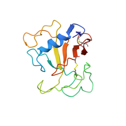
The deletion mutant Delta(7-22) of alpha-sarcin, unlike its wild-type protein counterpart, lacks the specific ability to degrade rRNA in intact ribosomes and exhibits an increased unspecific ribonuclease activity and decreased interaction with lipid vesicles. In trying to shed light on these differences, we report here on the three-dimensional structure of the Delta(7-22) alpha-sarcin mutant using NMR methods. We also evaluated its dynamic properties on the basis of theoretical models and measured its correlation time (6.2 nsec) by time-resolved fluorescence anisotropy. The global fold characteristic of ribotoxins is preserved in the mutant. The most significant differences with respect to the alpha-sarcin structure are concentrated in (1) loop 2, (2) loop 3, which adopts a new orientation, and (3) loop 5, which shows multiple conformations and an altered dynamics. The interactions between loop 5 and the N-terminal hairpin are lost in the mutant, producing increased solvent accessibility of the active-site residues. The degree of solvent exposure of the catalytic His 137 is similar to that shown by His 92 in RNase T1. Additionally, the calculated order parameters of residues belonging to loop 5 in the mutant correspond to an internal dynamic behavior more similar to RNase T1 than alpha-sarcin. On the other hand, changes in the relative orientation of loop 3 move the lysine-rich region 111-114, crucial for substrate recognition, away from the active site. All of the structural and dynamic data presented here reveal that the mutant is a hybrid of ribotoxins and noncytotoxic ribonucleases, consistent with its biological properties.
Organizational Affiliation:
Departamento de Espectroscopía y Estructura Molecular, Instituto de Química Física Rocasolano, Serrano 119, CSIC, 28006 Madrid, Spain.