1R17

Crystal Structure Analysis of S.epidermidis adhesin SdrG binding to Fibrinogen (adhesin-ligand complex)


Experimental Data Snapshot

  • Method: X-RAY DIFFRACTION
  • Resolution: 1.86 Å
  • R-Value Free: 0.221 
  • R-Value Work: 0.210 
  • R-Value Observed: 0.210 

wwPDB Validation   3D Report Full Report


This is version 1.3 of the entry. See complete history


Literature

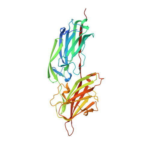
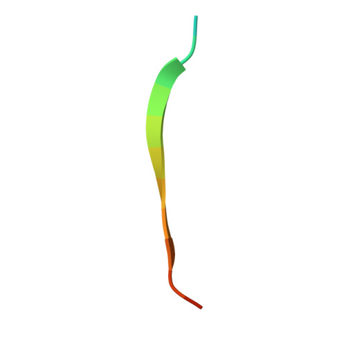
A "dock, lock and latch" Structural Model for a Staphylococcal Adhesin Binding to Fibrinogen

Ponnuraj, K.Bowden, M.G.Davis, S.Gurusiddappa, S.Moore, D.Choe, D.Xu, Y.Hook, M.Narayana, S.V.L.

(2003) Cell 115: 217-228

  • DOI: https://doi.org/10.1016/s0092-8674(03)00809-2
  • Primary Citation of Related Structures:  
    1R17, 1R19

  • PubMed Abstract: 

    Gram-positive pathogens such as staphylococci contain multiple cell wall-anchored proteins that serve as an interface between the microbe and its environment. Some of these proteins act as adhesins and mediate bacterial attachment to host tissues. SdrG is a cell wall-anchored adhesin from Staphylococcus epidermidis that binds to the Bbeta chain of human fibrinogen (Fg) and is necessary and sufficient for bacterial attachment to Fg-coated biomaterials. Here, we present the crystal structures of the ligand binding region of SdrG as an apoprotein and in complex with a synthetic peptide analogous to its binding site in Fg. Analysis of the crystal structures, along with mutational studies of both the protein and of the peptide, reveals that SdrG binds to its ligand with a dynamic "dock, lock, and latch" mechanism. We propose that this mechanism represents a general mode of ligand binding for structurally related cell wall-anchored proteins of gram-positive bacteria.


  • Organizational Affiliation

    School of Optometry and Center for Biophysical Sciences and Engineering, University of Alabama at Birmingham, Birmingham, AL 35294, USA.


Macromolecules
Find similar proteins by:  (by identity cutoff)  |  3D Structure
Entity ID: 1
MoleculeChains Sequence LengthOrganismDetailsImage
fibrinogen-binding protein SdrG
A, B
343Staphylococcus epidermidisMutation(s): 0 
Gene Names: SDRG
UniProt
Find proteins for Q9KI13 (Staphylococcus epidermidis)
Explore Q9KI13 
Go to UniProtKB:  Q9KI13
Entity Groups  
Sequence Clusters30% Identity50% Identity70% Identity90% Identity95% Identity100% Identity
UniProt GroupQ9KI13
Sequence Annotations
Expand
  • Reference Sequence

Find similar proteins by:  Sequence   |   3D Structure  

Entity ID: 2
MoleculeChains Sequence LengthOrganismDetailsImage
fibrinopeptide B
C, D
16N/AMutation(s): 0 
Entity Groups  
Sequence Clusters30% Identity50% Identity70% Identity90% Identity95% Identity100% Identity
Sequence Annotations
Expand
  • Reference Sequence
Experimental Data & Validation

Experimental Data

  • Method: X-RAY DIFFRACTION
  • Resolution: 1.86 Å
  • R-Value Free: 0.221 
  • R-Value Work: 0.210 
  • R-Value Observed: 0.210 
  • Space Group: P 21 21 21
Unit Cell:
Length ( Å )Angle ( ˚ )
a = 89.018α = 90
b = 89.483β = 90
c = 98.044γ = 90
Software Package:
Software NamePurpose
CNSrefinement
DENZOdata reduction
SCALEPACKdata scaling
SOLVEphasing
CNSphasing

Structure Validation

View Full Validation Report


Entry History 

Deposition Data

Revision History  (Full details and data files)

  • Version 1.0: 2003-10-28
    Type: Initial release
  • Version 1.1: 2008-04-29
    Changes: Version format compliance
  • Version 1.2: 2011-07-13
    Changes: Version format compliance
  • Version 1.3: 2024-02-14
    Changes: Data collection, Database references, Derived calculations