1QXM

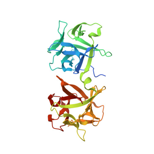
Crystal structure of a hemagglutinin component (HA1) from type C Clostridium botulinum


Experimental Data Snapshot

  • Method: X-RAY DIFFRACTION
  • Resolution: 1.70 Å
  • R-Value Free: 0.201 
  • R-Value Work: 0.177 
  • R-Value Observed: 0.177 

wwPDB Validation   3D Report Full Report


This is version 1.4 of the entry. See complete history


Literature

Structural analysis by X-ray crystallography and calorimetry of a haemagglutinin component (HA1) of the progenitor toxin from Clostridium botulinum.

Inoue, K.Sobhany, M.Transue, T.R.Oguma, K.Pedersen, L.C.Negishi, M.

(2003) Microbiology (N Y) 149: 3361-3370

  • DOI: https://doi.org/10.1099/mic.0.26586-0
  • Primary Citation of Related Structures:  
    1QXM

  • PubMed Abstract: 

    Botulism food poisoning is caused primarily by ingestion of the Clostridium botulinum neurotoxin (BoNT). The 1300 amino acid BoNT forms a progenitor toxin (PTX) that, when associated with a number of other proteins, increases its oral toxicity by protecting it from the low pH of the stomach and from intestinal proteases. One of these associated proteins, HA1, has also been suggested to be involved with internalization of the toxin into the bloodstream by binding to oligosaccharides lining the intestine. Here is reported the crystal structure of HA1 from type C Clostridium botulinum at a resolution of 1.7 Angstrom. The protein consists of two beta-trefoil domains and bears structural similarities to the lectin B-chain from the deadly plant toxin ricin. Based on structural comparison to the ricin B-chain lactose-binding sites, residues of type A HA1 were selected and mutated. The D263A and N285A mutants lost the ability to bind carbohydrates containing galactose moieties, implicating these residues in carbohydrate binding.


  • Organizational Affiliation

    Pharmacogenetic Section Laboratory of Reproductive and Developmental Toxicology, National Institute of Environmental Health Sciences, National Institutes of Health, Research Triangle Park, NC 27709, USA.


Macromolecules
Find similar proteins by:  (by identity cutoff)  |  3D Structure
Entity ID: 1
MoleculeChains Sequence LengthOrganismDetailsImage
HA1
A, B
300Clostridium botulinum D phageMutation(s): 0 
UniProt
Find proteins for P0DPR1 (Clostridium botulinum D phage)
Explore P0DPR1 
Go to UniProtKB:  P0DPR1
Entity Groups  
Sequence Clusters30% Identity50% Identity70% Identity90% Identity95% Identity100% Identity
UniProt GroupP0DPR1
Sequence Annotations
Expand
  • Reference Sequence
Experimental Data & Validation

Experimental Data

  • Method: X-RAY DIFFRACTION
  • Resolution: 1.70 Å
  • R-Value Free: 0.201 
  • R-Value Work: 0.177 
  • R-Value Observed: 0.177 
  • Space Group: C 1 2 1
Unit Cell:
Length ( Å )Angle ( ˚ )
a = 138.173α = 90
b = 62.332β = 104.53
c = 82.321γ = 90
Software Package:
Software NamePurpose
CNSrefinement
SCALEPACKdata scaling
MLPHAREphasing

Structure Validation

View Full Validation Report


Entry History 

Deposition Data

Revision History  (Full details and data files)

  • Version 1.0: 2004-01-20
    Type: Initial release
  • Version 1.1: 2008-04-29
    Changes: Version format compliance
  • Version 1.2: 2011-07-13
    Changes: Version format compliance
  • Version 1.3: 2017-10-11
    Changes: Refinement description
  • Version 1.4: 2024-02-14
    Changes: Data collection, Database references, Derived calculations