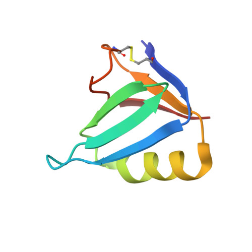
A Mutant Shiga-Like Toxin Iie Bound to its Receptor Gb(3): Structure of a Group II Shiga-Like Toxin with Altered Binding Specificity
Ling, H., Pannu, N.S., Boodhoo, A., Armstrong, G.D., Clark, C.G., Brunton, J.L., Read, R.J.(2000) Structure 8: 253
- PubMed: 10745005
- DOI: https://doi.org/10.1016/s0969-2126(00)00103-9
- Primary Citation of Related Structures:
1QOH, 2BOS - PubMed Abstract:
Shiga-like toxins (SLTs) are produced by the pathogenic strains of Escherichia coli that cause hemorrhagic colitis and hemolytic uremic syndrome. These diseases in humans are generally associated with group II family members (SLT-II and SLT-IIc), whereas SLT-IIe (pig edema toxin) is central to edema disease of swine. The pentameric B-subunit component of the majority of family members binds to the cell-surface glycolipid globotriaosyl ceramide (Gb(3)), but globotetraosyl ceramide (Gb(4)) is the preferred receptor for SLT-IIe. A double-mutant of the SLT-IIe B subunit that reverses two sequence differences from SLT-II (GT3; Gln65-->Glu, Lys67-->Gln, SLT-I numbering) has been shown to bind more strongly to Gb(3) than to Gb(4). To understand the molecular basis of receptor binding and specificity, we have determined the structure of the GT3 mutant B pentamer, both in complex with a Gb(3) analogue (2.0 A resolution; R = 0.155, R(free) = 0.194) and in its native form (2.35 A resolution; R = 0.187, R(free) = 0.232). These are the first structures of a member of the medically important group II Shiga-like toxins to be reported. The structures confirm the previous observation of multiple binding sites on each SLT monomer, although binding site 3 is not occupied in the GT3 structure. Analysis of the binding properties of mutants suggests that site 3 is a secondary Gb(4)-binding site. The two mutated residues are located appropriately to interact with the extra betaGalNAc residue on Gb(4). Differences in the binding sites provide a molecular basis for understanding the tissue specificities and pathogenic mechanisms of members of the SLT family.
Organizational Affiliation:
Department of Biochemistry, University of Alberta, Edmonton, T6G 2H7, Canada.


