1QKS

CYTOCHROME CD1 NITRITE REDUCTASE, OXIDISED FORM


Experimental Data Snapshot

  • Method: X-RAY DIFFRACTION
  • Resolution: 1.28 Å
  • R-Value Free: 0.200 
  • R-Value Work: 0.185 
  • R-Value Observed: 0.185 

wwPDB Validation   3D Report Full Report


This is version 1.4 of the entry. See complete history


Literature

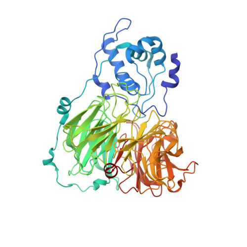
The anatomy of a bifunctional enzyme: structural basis for reduction of oxygen to water and synthesis of nitric oxide by cytochrome cd1.

Fulop, V.Moir, J.W.Ferguson, S.J.Hajdu, J.

(1995) Cell 81: 369-377

  • DOI: https://doi.org/10.1016/0092-8674(95)90390-9
  • Primary Citation of Related Structures:  
    1QKS

  • PubMed Abstract: 

    Cytochrome cd1-nitrite reductase is a bifunctional enzyme that catalyzes the one-electron reduction of nitrite to nitric oxide and the four-electron reduction of oxygen to water. The 1.55 A crystal structure of the dimeric enzyme from Thiosphaera pantotropha is reported here. The protein was sequenced from the X-ray structure. Each subunit contains a covalent c heme with two axial His ligands (His-17, His-69) and a unique noncovalent d1 heme ligated by Tyr-25 and His-200. The d1 heme is the mononuclear iron center where both oxygen and nitrite reduction take place. The two types of heme are located in separate domains whose arrangement suggests a mechanism requiring domain movement during catalysis.


  • Organizational Affiliation

    Laboratory of Molecular Biophysics, University of Oxford, England.


Macromolecules
Find similar proteins by:  (by identity cutoff)  |  3D Structure
Entity ID: 1
MoleculeChains Sequence LengthOrganismDetailsImage
CYTOCHROME CD1 NITRITE REDUCTASE
A, B
567Paracoccus pantotrophusMutation(s): 0 
UniProt
Find proteins for P72181 (Paracoccus pantotrophus)
Explore P72181 
Go to UniProtKB:  P72181
Entity Groups  
Sequence Clusters30% Identity50% Identity70% Identity90% Identity95% Identity100% Identity
UniProt GroupP72181
Sequence Annotations
Expand
  • Reference Sequence
Small Molecules
Ligands 4 Unique
IDChains Name / Formula / InChI Key2D Diagram3D Interactions
DHE
Query on DHE

Download Ideal Coordinates CCD File 
D [auth A],
L [auth B]
HEME D
C34 H32 Fe N4 O10
XLQCGNUTSJTZNF-YDXXJHAFSA-L
HEC
Query on HEC

Download Ideal Coordinates CCD File 
C [auth A],
K [auth B]
HEME C
C34 H34 Fe N4 O4
HXQIYSLZKNYNMH-LJNAALQVSA-N
SO4
Query on SO4

Download Ideal Coordinates CCD File 
I [auth A]
J [auth A]
P [auth B]
Q [auth B]
R [auth B]
I [auth A],
J [auth A],
P [auth B],
Q [auth B],
R [auth B],
S [auth B]
SULFATE ION
O4 S
QAOWNCQODCNURD-UHFFFAOYSA-L
GOL
Query on GOL

Download Ideal Coordinates CCD File 
E [auth A]
F [auth A]
G [auth A]
H [auth A]
M [auth B]
E [auth A],
F [auth A],
G [auth A],
H [auth A],
M [auth B],
N [auth B],
O [auth B]
GLYCEROL
C3 H8 O3
PEDCQBHIVMGVHV-UHFFFAOYSA-N
Experimental Data & Validation

Experimental Data

  • Method: X-RAY DIFFRACTION
  • Resolution: 1.28 Å
  • R-Value Free: 0.200 
  • R-Value Work: 0.185 
  • R-Value Observed: 0.185 
  • Space Group: P 1 21 1
Unit Cell:
Length ( Å )Angle ( ˚ )
a = 106.4α = 90
b = 60.6β = 112.3
c = 100.2γ = 90
Software Package:
Software NamePurpose
X-PLORrefinement
DENZOdata reduction
SCALEPACKdata scaling
X-PLORphasing

Structure Validation

View Full Validation Report


Entry History 

Deposition Data

  • Released Date: 1999-08-18 
  • Deposition Author(s): Fulop, V.

Revision History  (Full details and data files)

  • Version 1.0: 1999-08-18
    Type: Initial release
  • Version 1.1: 2011-05-08
    Changes: Version format compliance
  • Version 1.2: 2011-07-13
    Changes: Version format compliance
  • Version 1.3: 2019-01-16
    Changes: Data collection, Database references
  • Version 1.4: 2019-10-09
    Changes: Data collection, Database references, Derived calculations