1QD9

Bacillus subtilis YABJ


Experimental Data Snapshot

  • Method: X-RAY DIFFRACTION
  • Resolution: 1.70 Å
  • R-Value Free: 0.197 
  • R-Value Work: 0.166 
  • R-Value Observed: 0.166 

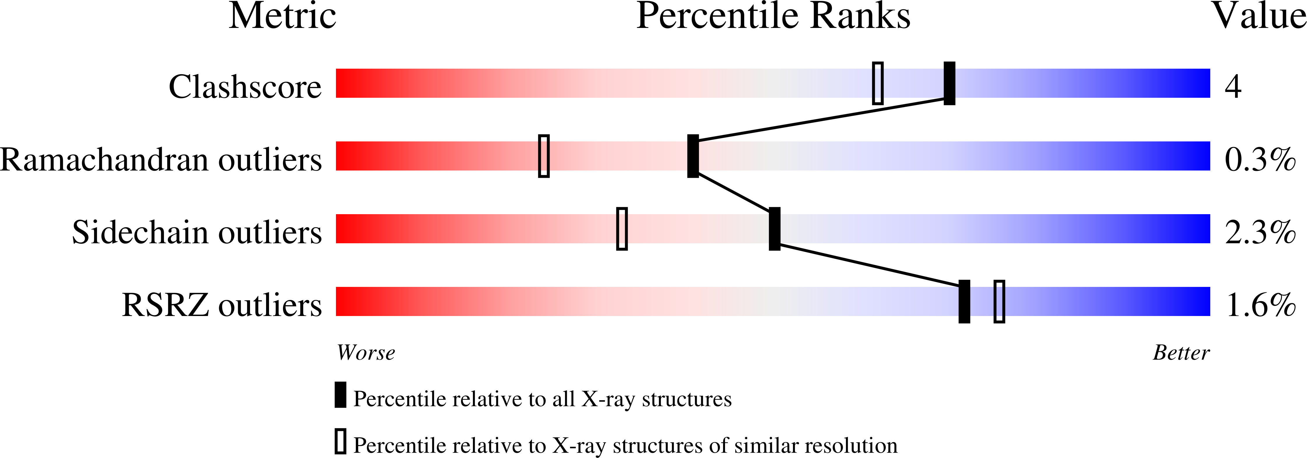
wwPDB Validation   3D Report Full Report


This is version 1.3 of the entry. See complete history


Literature

Crystal structure of Bacillus subtilis YabJ, a purine regulatory protein and member of the highly conserved YjgF family.

Sinha, S.Rappu, P.Lange, S.C.Mantsala, P.Zalkin, H.Smith, J.L.

(1999) Proc Natl Acad Sci U S A 96: 13074-13079

  • DOI: https://doi.org/10.1073/pnas.96.23.13074
  • Primary Citation of Related Structures:  
    1QD9

  • PubMed Abstract: 

    The yabJ gene in Bacillus subtilis is required for adenine-mediated repression of purine biosynthetic genes in vivo and codes for an acid-soluble, 14-kDa protein. The molecular mechanism of YabJ is unknown. YabJ is a member of a large, widely distributed family of proteins of unknown biochemical function. The 1.7-A crystal structure of YabJ reveals a trimeric organization with extensive buried hydrophobic surface and an internal water-filled cavity. The most important finding in the structure is a deep, narrow cleft between subunits lined with nine side chains that are invariant among the 25 most similar homologs. This conserved site is proposed to be a binding or catalytic site for a ligand or substrate that is common to YabJ and other members of the YER057c/YjgF/UK114 family of proteins.


  • Organizational Affiliation

    Department of Biological Sciences, Purdue University, West Lafayette, IN 47907, USA.


Macromolecules
Find similar proteins by:  (by identity cutoff)  |  3D Structure
Entity ID: 1
MoleculeChains Sequence LengthOrganismDetailsImage
PURINE REGULATORY PROTEIN YABJ
A, B, C
124Bacillus subtilisMutation(s): 0 
UniProt
Find proteins for P37552 (Bacillus subtilis (strain 168))
Explore P37552 
Go to UniProtKB:  P37552
Entity Groups  
Sequence Clusters30% Identity50% Identity70% Identity90% Identity95% Identity100% Identity
UniProt GroupP37552
Sequence Annotations
Expand
  • Reference Sequence
Small Molecules
Ligands 3 Unique
IDChains Name / Formula / InChI Key2D Diagram3D Interactions
EMC
Query on EMC

Download Ideal Coordinates CCD File 
G [auth B],
K [auth C]
ETHYL MERCURY ION
C2 H5 Hg
MJOUBOKSWBMNGQ-UHFFFAOYSA-N
HG
Query on HG

Download Ideal Coordinates CCD File 
D [auth A]MERCURY (II) ION
Hg
BQPIGGFYSBELGY-UHFFFAOYSA-N
ACY
Query on ACY

Download Ideal Coordinates CCD File 
E [auth A]
F [auth A]
H [auth B]
I [auth B]
J [auth B]
E [auth A],
F [auth A],
H [auth B],
I [auth B],
J [auth B],
L [auth C]
ACETIC ACID
C2 H4 O2
QTBSBXVTEAMEQO-UHFFFAOYSA-N
Experimental Data & Validation

Experimental Data

  • Method: X-RAY DIFFRACTION
  • Resolution: 1.70 Å
  • R-Value Free: 0.197 
  • R-Value Work: 0.166 
  • R-Value Observed: 0.166 
  • Space Group: P 65
Unit Cell:
Length ( Å )Angle ( ˚ )
a = 53.35α = 90
b = 53.35β = 90
c = 204.96γ = 120
Software Package:
Software NamePurpose
DENZOdata reduction
SCALEPACKdata scaling
MLPHAREphasing
CNSrefinement

Structure Validation

View Full Validation Report


Entry History 

Deposition Data

Revision History  (Full details and data files)

  • Version 1.0: 1999-11-26
    Type: Initial release
  • Version 1.1: 2008-04-27
    Changes: Version format compliance
  • Version 1.2: 2011-07-13
    Changes: Version format compliance
  • Version 1.3: 2024-02-14
    Changes: Data collection, Database references, Derived calculations