1Q4D

S65T Q80R T203C Green Fluorescent Protein (GFP) pH 5.5


Experimental Data Snapshot

  • Method: X-RAY DIFFRACTION
  • Resolution: 1.58 Å
  • R-Value Free: 0.233 
  • R-Value Work: 0.208 
  • R-Value Observed: 0.208 

wwPDB Validation   3D Report Full Report


This is version 1.5 of the entry. See complete history


Literature

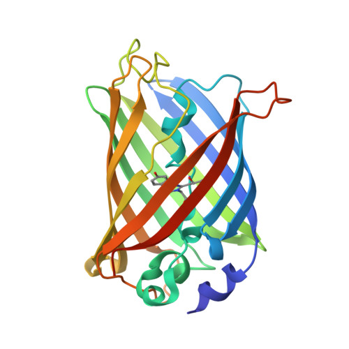
Local complexity of amino acid interactions in a protein core.

Jain, R.K.Ranganathan, R.

(2004) Proc Natl Acad Sci U S A 101: 111-116

  • DOI: https://doi.org/10.1073/pnas.2534352100
  • Primary Citation of Related Structures:  
    1Q4A, 1Q4B, 1Q4C, 1Q4D, 1Q4E, 1Q73

  • PubMed Abstract: 

    Atomic resolution structures of proteins indicate that the core is typically well packed, suggesting a densely connected network of interactions between amino acid residues. The combinatorial complexity of energetic interactions in such a network could be enormous, a problem that limits our ability to relate structure and function. Here, we report a case study of the complexity of amino acid interactions in a localized region within the core of the GFP, a particularly stable and tightly packed molecule. Mutations at three sites within the chromophore-binding pocket display an overlapping pattern of conformational change and are thermodynamically coupled, seemingly consistent with the dense network model. However, crystallographic and energetic analyses of coupling between mutations paint a different picture; pairs of mutations couple through independent "hotspots" in the region of structural overlap. The data indicate that, even in highly stable proteins, the core contains sufficient plasticity in packing to uncouple high-order energetic interactions of residues, a property that is likely general in proteins.


  • Organizational Affiliation

    Howard Hughes Medical Institute and Department of Pharmacology, University of Texas Southwestern Medical Center, 5323 Harry Hines Boulevard, Dallas, TX 75390-9050, USA.


Macromolecules
Find similar proteins by:  (by identity cutoff)  |  3D Structure
Entity ID: 1
MoleculeChains Sequence LengthOrganismDetailsImage
Green Fluorescent Protein236Aequorea victoriaMutation(s): 3 
UniProt
Find proteins for P42212 (Aequorea victoria)
Explore P42212 
Go to UniProtKB:  P42212
Entity Groups  
Sequence Clusters30% Identity50% Identity70% Identity90% Identity95% Identity100% Identity
UniProt GroupP42212
Sequence Annotations
Expand
  • Reference Sequence
Small Molecules
Modified Residues  1 Unique
IDChains TypeFormula2D DiagramParent
CRO
Query on CRO
A
L-PEPTIDE LINKINGC15 H17 N3 O5THR, TYR, GLY
Experimental Data & Validation

Experimental Data

  • Method: X-RAY DIFFRACTION
  • Resolution: 1.58 Å
  • R-Value Free: 0.233 
  • R-Value Work: 0.208 
  • R-Value Observed: 0.208 
  • Space Group: P 21 21 21
Unit Cell:
Length ( Å )Angle ( ˚ )
a = 50.969α = 90
b = 62.333β = 90
c = 68.554γ = 90
Software Package:
Software NamePurpose
DENZOdata reduction
SCALEPACKdata scaling
CNSrefinement
CNSphasing

Structure Validation

View Full Validation Report


Entry History 

Deposition Data

Revision History  (Full details and data files)

  • Version 1.0: 2004-02-03
    Type: Initial release
  • Version 1.1: 2008-04-29
    Changes: Version format compliance
  • Version 1.2: 2011-07-13
    Changes: Version format compliance
  • Version 1.3: 2021-11-10
    Changes: Database references, Derived calculations
  • Version 1.4: 2023-10-25
    Changes: Data collection, Refinement description
  • Version 1.5: 2023-11-15
    Changes: Data collection