1Q0X

Anti-morphine Antibody 9B1 Unliganded Form


Experimental Data Snapshot

  • Method: X-RAY DIFFRACTION
  • Resolution: 1.60 Å
  • R-Value Free: 0.251 
  • R-Value Work: 0.216 
  • R-Value Observed: 0.216 

wwPDB Validation   3D Report Full Report


This is version 1.4 of the entry. See complete history


Literature

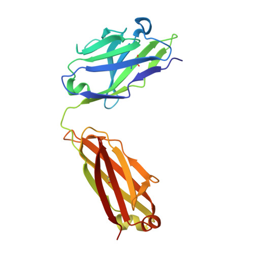
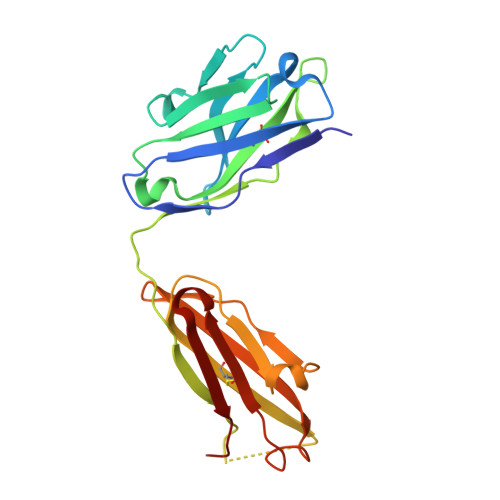
Anchoring a cationic ligand: the structure of the Fab fragment of the anti-morphine antibody 9B1 and its complex with morphine

Pozharski, E.Wilson, M.A.Hewagama, A.Shanafelt, A.B.Petsko, G.Ringe, D.

(2004) J Mol Biol 337: 691-697

  • DOI: https://doi.org/10.1016/j.jmb.2003.12.084
  • Primary Citation of Related Structures:  
    1Q0X, 1Q0Y

  • PubMed Abstract: 

    The crystal structures of an anti-morphine antibody 9B1 (to 1.6A resolution) and its complex with morphine (to 2.0 A resolution) are reported. The morphine-binding site is described as a shallow depression on the protein surface, an unusual topology for a high-affinity ( Ka approximately 10(9) M(-1)) antibody against a small antigen. The polar part of the ligand is exposed to solvent, and the cationic nitrogen atom of the morphine molecule is anchored at the bottom of the binding site by a salt-bridge to a glutamate side-chain. Additional affinity is provided by a double cation-pi interaction with two tryptophan residues. Comparison of the morphine complex with the structure of the free Fab shows that a domain closure occurs upon binding of the ligand.


  • Organizational Affiliation

    Rosenstiel Basic Medical Sciences Research Center, Brandeis University, 415 South Street, Waltham, MA 02454, USA.


Macromolecules
Find similar proteins by:  (by identity cutoff)  |  3D Structure
Entity ID: 1
MoleculeChains Sequence LengthOrganismDetailsImage
Fab 9B1, light chainA [auth L]212Mus musculusMutation(s): 0 
UniProt
Find proteins for P01723 (Mus musculus)
Explore P01723 
Go to UniProtKB:  P01723
Entity Groups  
Sequence Clusters30% Identity50% Identity70% Identity90% Identity95% Identity100% Identity
UniProt GroupP01723
Sequence Annotations
Expand
  • Reference Sequence
Find similar proteins by:  (by identity cutoff)  |  3D Structure
Entity ID: 2
MoleculeChains Sequence LengthOrganismDetailsImage
Fab 9B1, heavy chainB [auth H]221Mus musculusMutation(s): 0 
UniProt
Find proteins for Q9D9B8 (Mus musculus)
Explore Q9D9B8 
Go to UniProtKB:  Q9D9B8
Entity Groups  
Sequence Clusters30% Identity50% Identity70% Identity90% Identity95% Identity100% Identity
UniProt GroupQ9D9B8
Sequence Annotations
Expand
  • Reference Sequence
Experimental Data & Validation

Experimental Data

  • Method: X-RAY DIFFRACTION
  • Resolution: 1.60 Å
  • R-Value Free: 0.251 
  • R-Value Work: 0.216 
  • R-Value Observed: 0.216 
  • Space Group: C 1 2 1
Unit Cell:
Length ( Å )Angle ( ˚ )
a = 56.514α = 90
b = 59.924β = 92.36
c = 114.706γ = 90
Software Package:
Software NamePurpose
DENZOdata reduction
SCALEPACKdata scaling
AMoREphasing
CNSrefinement

Structure Validation

View Full Validation Report


Entry History 

Deposition Data

Revision History  (Full details and data files)

  • Version 1.0: 2004-04-20
    Type: Initial release
  • Version 1.1: 2008-04-29
    Changes: Version format compliance
  • Version 1.2: 2011-07-13
    Changes: Version format compliance
  • Version 1.3: 2011-11-16
    Changes: Atomic model
  • Version 1.4: 2023-08-16
    Changes: Data collection, Database references, Derived calculations, Refinement description