1PYT

TERNARY COMPLEX OF PROCARBOXYPEPTIDASE A, PROPROTEINASE E, AND CHYMOTRYPSINOGEN C


Experimental Data Snapshot

  • Method: X-RAY DIFFRACTION
  • Resolution: 2.35 Å
  • R-Value Free: 0.283 
  • R-Value Work: 0.192 
  • R-Value Observed: 0.192 

wwPDB Validation   3D Report Full Report


This is version 1.2 of the entry. See complete history


Literature

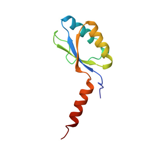
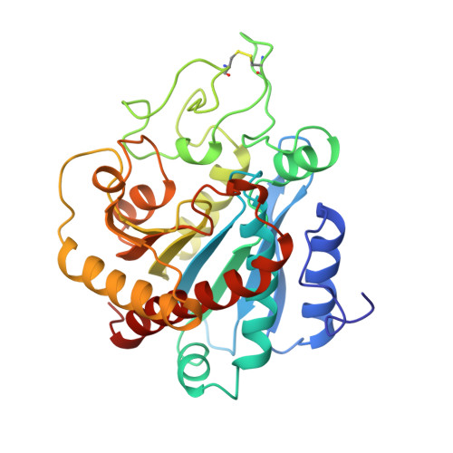
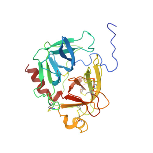
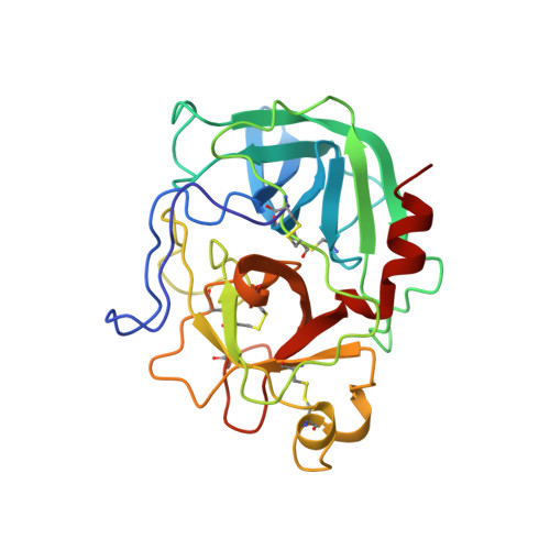
The three-dimensional structure of the native ternary complex of bovine pancreatic procarboxypeptidase A with proproteinase E and chymotrypsinogen C.

Gomis-Ruth, F.X.Gomez, M.Bode, W.Huber, R.Aviles, F.X.

(1995) EMBO J 14: 4387-4394

  • DOI: https://doi.org/10.1002/j.1460-2075.1995.tb00117.x
  • Primary Citation of Related Structures:  
    1PYT

  • PubMed Abstract: 

    The metalloexozymogen procarboxypeptidase A is mainly secreted in ruminants as a ternary complex with zymogens of two serine endoproteinases, chymotrypsinogen C and proproteinase E. The bovine complex has been crystallized, and its molecular structure analysed and refined at 2.6 A resolution to an R factor of 0.198. In this heterotrimer, the activation segment of procarboxypeptidase A essentially clamps the other two subunits, which shield the activation sites of the former from tryptic attack. In contrast, the propeptides of both serine proproteinases are freely accessible to trypsin. This arrangement explains the sequential and delayed activation of the constituent zymogens. Procarboxypeptidase A is virtually identical to the homologous monomeric porcine form. Chymotrypsinogen C displays structural features characteristic for chymotrypsins as well as elastases, except for its activation domain; similar to bovine chymotrypsinogen A, its binding site is not properly formed, while its surface located activation segment is disordered. The proproteinase E structure is fully ordered and strikingly similar to active porcine elastase; its specificity pocket is occluded, while the activation segment is fixed to the molecular surface. This first structure of a native zymogen from the proteinase E/elastase family does not fundamentally differ from the serine proproteinases known so far.


  • Organizational Affiliation

    Institut de Biologia Fonamental, Universitat Autònoma de Barcelona, Bellaterra, Spain.


Macromolecules
Find similar proteins by:  (by identity cutoff)  |  3D Structure
Entity ID: 1
MoleculeChains Sequence LengthOrganismDetailsImage
PROCARBOXYPEPTIDASE A94Bos taurusMutation(s): 0 
EC: 3.4.17.1
UniProt
Find proteins for P00730 (Bos taurus)
Explore P00730 
Go to UniProtKB:  P00730
Entity Groups  
Sequence Clusters30% Identity50% Identity70% Identity90% Identity95% Identity100% Identity
UniProt GroupP00730
Sequence Annotations
Expand
  • Reference Sequence
Find similar proteins by:  (by identity cutoff)  |  3D Structure
Entity ID: 2
MoleculeChains Sequence LengthOrganismDetailsImage
PROCARBOXYPEPTIDASE A309Bos taurusMutation(s): 0 
EC: 3.4.17.1
UniProt
Find proteins for P00730 (Bos taurus)
Explore P00730 
Go to UniProtKB:  P00730
Entity Groups  
Sequence Clusters30% Identity50% Identity70% Identity90% Identity95% Identity100% Identity
UniProt GroupP00730
Sequence Annotations
Expand
  • Reference Sequence
Find similar proteins by:  (by identity cutoff)  |  3D Structure
Entity ID: 3
MoleculeChains Sequence LengthOrganismDetailsImage
PROPROTEINASE E253Bos taurusMutation(s): 0 
UniProt
Find proteins for P05805 (Bos taurus)
Explore P05805 
Go to UniProtKB:  P05805
Entity Groups  
Sequence Clusters30% Identity50% Identity70% Identity90% Identity95% Identity100% Identity
UniProt GroupP05805
Sequence Annotations
Expand
  • Reference Sequence
Find similar proteins by:  (by identity cutoff)  |  3D Structure
Entity ID: 4
MoleculeChains Sequence LengthOrganismDetailsImage
CHYMOTRYPSINOGEN C251Bos taurusMutation(s): 0 
UniProt
Find proteins for Q7M3E1 (Bos taurus)
Explore Q7M3E1 
Go to UniProtKB:  Q7M3E1
Entity Groups  
Sequence Clusters30% Identity50% Identity70% Identity90% Identity95% Identity100% Identity
UniProt GroupQ7M3E1
Sequence Annotations
Expand
  • Reference Sequence
Small Molecules
Ligands 2 Unique
IDChains Name / Formula / InChI Key2D Diagram3D Interactions
ZN
Query on ZN

Download Ideal Coordinates CCD File 
E [auth B]ZINC ION
Zn
PTFCDOFLOPIGGS-UHFFFAOYSA-N
CA
Query on CA

Download Ideal Coordinates CCD File 
F [auth C]CALCIUM ION
Ca
BHPQYMZQTOCNFJ-UHFFFAOYSA-N
Experimental Data & Validation

Experimental Data

  • Method: X-RAY DIFFRACTION
  • Resolution: 2.35 Å
  • R-Value Free: 0.283 
  • R-Value Work: 0.192 
  • R-Value Observed: 0.192 
  • Space Group: H 3
Unit Cell:
Length ( Å )Angle ( ˚ )
a = 188.5α = 90
b = 188.5β = 90
c = 82.5γ = 120
Software Package:
Software NamePurpose
MOSFLMdata reduction
X-PLORmodel building
X-PLORrefinement
X-PLORphasing

Structure Validation

View Full Validation Report


Entry History 

Deposition Data

Revision History  (Full details and data files)

  • Version 1.0: 1997-01-27
    Type: Initial release
  • Version 1.1: 2008-03-03
    Changes: Version format compliance
  • Version 1.2: 2011-07-13
    Changes: Version format compliance