1PSK

THE CRYSTAL STRUCTURE OF AN FAB FRAGMENT THAT BINDS TO THE MELANOMA-ASSOCIATED GD2 GANGLIOSIDE


Experimental Data Snapshot

  • Method: X-RAY DIFFRACTION
  • Resolution: 2.80 Å
  • R-Value Free: 0.326 
  • R-Value Work: 0.204 
  • R-Value Observed: 0.204 

wwPDB Validation   3D Report Full Report


This is version 1.3 of the entry. See complete history


Literature

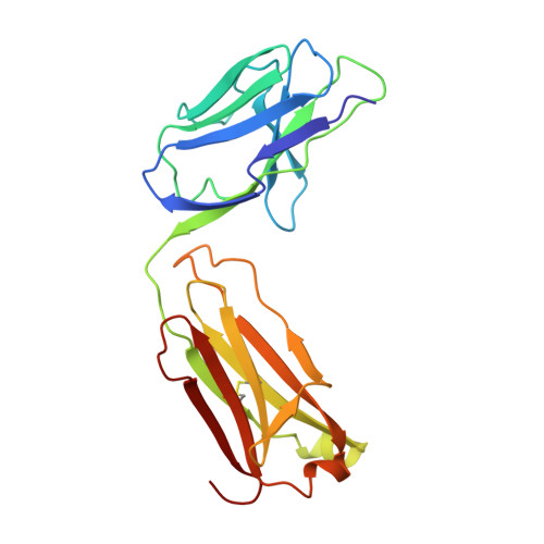
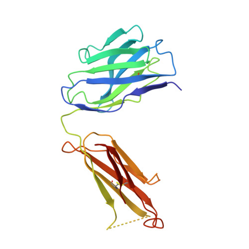
The crystal structure of a Fab fragment to the melanoma-associated GD2 ganglioside.

Pichla, S.L.Murali, R.Burnett, R.M.

(1997) J Struct Biol 119: 6-16

  • DOI: https://doi.org/10.1006/jsbi.1997.3857
  • Primary Citation of Related Structures:  
    1PSK

  • PubMed Abstract: 

    The GD2 ganglioside is a cell-surface component that appears on the surface of metastatic melanoma cells and is a marker for the progression of the disease. The ME36.1 monoclonal antibody binds to the GD2 ganglioside and has shown potential as a therapeutic antibody. ME36.1 is a possible alternative therapy to radiation, which is often ineffective in late-stage melanoma. The crystal structure of the Fab fragment of ME36.1 has been determined using molecular replacement and refined to an R factor of 20.4% at 2.8 A resolution. The model has good geometry with root-mean-square deviations of 0.008 A from ideal bond lengths and 1.7 degrees from ideal bond angles. The crystal structure of the ME36.1 Fab shows that its complementarity determining region forms a groove-shaped binding site rather than the pocket-type observed in other sugar binding Fabs. Molecular modeling has placed a four-residue sugar, representative of GD2, in the antigen binding site. The GD2 sugar moiety is stabilized by a network of hydrogen bonds that define the specificity of ME36.1 toward its antigen.


  • Organizational Affiliation

    Wistar Institute, Philadelphia, Pennsylvania 19104, USA.


Macromolecules
Find similar proteins by:  (by identity cutoff)  |  3D Structure
Entity ID: 1
MoleculeChains Sequence LengthOrganismDetailsImage
ANTIBODYA [auth L]213Mus musculusMutation(s): 0 
UniProt
Find proteins for P01837 (Mus musculus)
Explore P01837 
Go to UniProtKB:  P01837
Entity Groups  
Sequence Clusters30% Identity50% Identity70% Identity90% Identity95% Identity100% Identity
UniProt GroupP01837
Sequence Annotations
Expand
  • Reference Sequence
Find similar proteins by:  (by identity cutoff)  |  3D Structure
Entity ID: 2
MoleculeChains Sequence LengthOrganismDetailsImage
ANTIBODYB [auth H]209Mus musculusMutation(s): 0 
Entity Groups  
Sequence Clusters30% Identity50% Identity70% Identity90% Identity95% Identity100% Identity
Sequence Annotations
Expand
  • Reference Sequence
Experimental Data & Validation

Experimental Data

  • Method: X-RAY DIFFRACTION
  • Resolution: 2.80 Å
  • R-Value Free: 0.326 
  • R-Value Work: 0.204 
  • R-Value Observed: 0.204 
  • Space Group: P 1 21 1
Unit Cell:
Length ( Å )Angle ( ˚ )
a = 37.6α = 90
b = 94.1β = 101
c = 67.4γ = 90
Software Package:
Software NamePurpose
XDSdata scaling
XSCALEdata scaling
X-PLORmodel building
X-PLORrefinement
XDSdata reduction
X-PLORphasing

Structure Validation

View Full Validation Report


Entry History 

Deposition Data

Revision History  (Full details and data files)

  • Version 1.0: 1997-09-04
    Type: Initial release
  • Version 1.1: 2008-03-24
    Changes: Version format compliance
  • Version 1.2: 2011-07-13
    Changes: Version format compliance
  • Version 1.3: 2023-08-09
    Changes: Database references, Refinement description