1PNR

PURINE REPRESSOR-HYPOXANTHINE-PURF-OPERATOR COMPLEX


Experimental Data Snapshot

  • Method: X-RAY DIFFRACTION
  • Resolution: 2.70 Å
  • R-Value Observed: 0.171 

wwPDB Validation   3D Report Full Report


This is version 1.4 of the entry. See complete history


Literature

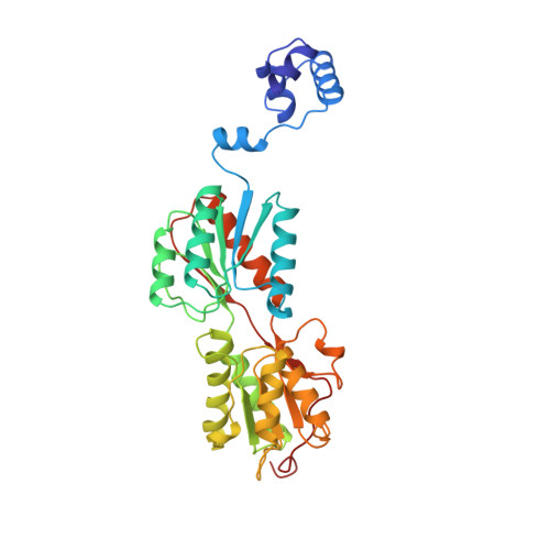
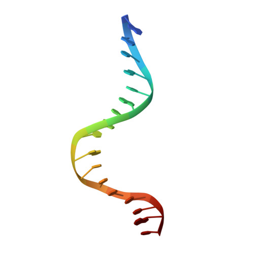
Crystal structure of LacI member, PurR, bound to DNA: minor groove binding by alpha helices.

Schumacher, M.A.Choi, K.Y.Zalkin, H.Brennan, R.G.

(1994) Science 266: 763-770

  • DOI: https://doi.org/10.1126/science.7973627
  • Primary Citation of Related Structures:  
    1PNR, 2PUA, 2PUB, 2PUC, 2PUD

  • PubMed Abstract: 

    The three-dimensional structure of a ternary complex of the purine repressor, PurR, bound to both its corepressor, hypoxanthine, and the 16-base pair purF operator site has been solved at 2.7 A resolution by x-ray crystallography. The bipartite structure of PurR consists of an amino-terminal DNA-binding domain and a larger carboxyl-terminal corepressor binding and dimerization domain that is similar to that of the bacterial periplasmic binding proteins. The DNA-binding domain contains a helix-turn-helix motif that makes base-specific contacts in the major groove of the DNA. Base contacts are also made by residues of symmetry-related alpha helices, the "hinge" helices, which bind deeply in the minor groove. Critical to hinge helix-minor groove binding is the intercalation of the side chains of Leu54 and its symmetry-related mate, Leu54', into the central CpG-base pair step. These residues thereby act as "leucine levers" to pry open the minor groove and kink the purF operator by 45 degrees.


  • Organizational Affiliation

    Department of Biochemistry and Molecular Biology, Oregon Health Sciences University, Portland 97201-3098.


Macromolecules

Find similar proteins by:  (by identity cutoff)  |  3D Structure
Entity ID: 2
MoleculeChains Sequence LengthOrganismDetailsImage
PROTEIN (PURINE REPRESSOR)B [auth A]340Escherichia coliMutation(s): 0 
UniProt
Find proteins for P0ACP7 (Escherichia coli (strain K12))
Explore P0ACP7 
Go to UniProtKB:  P0ACP7
Entity Groups  
Sequence Clusters30% Identity50% Identity70% Identity90% Identity95% Identity100% Identity
UniProt GroupP0ACP7
Sequence Annotations
Expand
  • Reference Sequence

Find similar nucleic acids by:  Sequence   |   3D Structure  

Entity ID: 1
MoleculeChains LengthOrganismImage
DNA (5'-D(*AP*AP*CP*GP*AP*AP*AP*AP*CP*GP*TP*TP*TP*TP*CP*GP*T )-3')A [auth B]17N/A
Sequence Annotations
Expand
  • Reference Sequence
Small Molecules
Ligands 1 Unique
IDChains Name / Formula / InChI Key2D Diagram3D Interactions
HPA
Query on HPA

Download Ideal Coordinates CCD File 
C [auth A]HYPOXANTHINE
C5 H4 N4 O
FDGQSTZJBFJUBT-UHFFFAOYSA-N
Experimental Data & Validation

Experimental Data

  • Method: X-RAY DIFFRACTION
  • Resolution: 2.70 Å
  • R-Value Observed: 0.171 
  • Space Group: C 2 2 21
Unit Cell:
Length ( Å )Angle ( ˚ )
a = 175.85α = 90
b = 94.79β = 90
c = 81.84γ = 90
Software Package:
Software NamePurpose
TNTrefinement

Structure Validation

View Full Validation Report


Entry History 

Deposition Data

Revision History  (Full details and data files)

  • Version 1.0: 1995-11-20
    Type: Initial release
  • Version 1.1: 2008-05-22
    Changes: Version format compliance
  • Version 1.2: 2011-07-13
    Changes: Version format compliance
  • Version 1.3: 2018-04-04
    Changes: Data collection
  • Version 1.4: 2024-02-14
    Changes: Data collection, Database references, Derived calculations