1PK1

Hetero SAM domain structure of Ph and Scm.


Experimental Data Snapshot

  • Method: X-RAY DIFFRACTION
  • Resolution: 1.80 Å
  • R-Value Free: 0.249 
  • R-Value Work: 0.233 
  • R-Value Observed: 0.233 

wwPDB Validation   3D Report Full Report


This is version 1.4 of the entry. See complete history


Literature

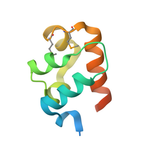
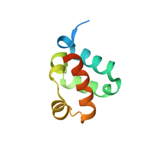
Structural organization of a Sex-comb-on-midleg/polyhomeotic copolymer.

Kim, C.A.Sawaya, M.R.Cascio, D.Kim, W.Bowie, J.U.

(2005) J Biol Chem 280: 27769-27775

  • DOI: https://doi.org/10.1074/jbc.M503055200
  • Primary Citation of Related Structures:  
    1PK1, 1PK3

  • PubMed Abstract: 

    The polycomb group proteins are required for the stable maintenance of gene repression patterns established during development. They function as part of large multiprotein complexes created via a multitude of protein-protein interaction domains. Here we examine the interaction between the SAM domains of the polycomb group proteins polyhomeotic (Ph) and Sex-comb-on-midleg (Scm). Previously we showed that Ph-SAM polymerizes as a helical structure. We find that Scm-SAM also polymerizes, and a crystal structure reveals an architecture similar to the Ph-SAM polymer. These results suggest that Ph-SAM and Scm-SAM form a copolymer. Binding affinity measurements between Scm-SAM and Ph-SAM subunits in different orientations indicate a preference for the formation of a single junction copolymer. To provide a model of the copolymer, we determined the structure of the Ph-SAM/Scm-SAM junction. Similar binding modes are observed in both homo- and heterocomplex formation with minimal change in helix axis direction at the polymer joint. The copolymer model suggests that polymeric Scm complexes could extend beyond the local domains of polymeric Ph complexes on chromatin, possibly playing a role in long range repression.


  • Organizational Affiliation

    Department of Chemistry and Biochemistry, University of California, Los Angeles, California 90095, USA.


Macromolecules
Find similar proteins by:  (by identity cutoff)  |  3D Structure
Entity ID: 1
MoleculeChains Sequence LengthOrganismDetailsImage
Polyhomeotic-proximal chromatin protein
A, C
89Drosophila melanogasterMutation(s): 3 
Gene Names: PH-P
UniProt
Find proteins for P39769 (Drosophila melanogaster)
Explore P39769 
Go to UniProtKB:  P39769
Entity Groups  
Sequence Clusters30% Identity50% Identity70% Identity90% Identity95% Identity100% Identity
UniProt GroupP39769
Sequence Annotations
Expand
  • Reference Sequence
Find similar proteins by:  (by identity cutoff)  |  3D Structure
Entity ID: 2
MoleculeChains Sequence LengthOrganismDetailsImage
Sex comb on midleg CG9495-PA
B, D
89Drosophila melanogasterMutation(s): 2 
Gene Names: Sex Comb on Midleg
UniProt
Find proteins for Q9VHA0 (Drosophila melanogaster)
Explore Q9VHA0 
Go to UniProtKB:  Q9VHA0
Entity Groups  
Sequence Clusters30% Identity50% Identity70% Identity90% Identity95% Identity100% Identity
UniProt GroupQ9VHA0
Sequence Annotations
Expand
  • Reference Sequence
Small Molecules
Modified Residues  1 Unique
IDChains TypeFormula2D DiagramParent
MSE
Query on MSE
A, C
L-PEPTIDE LINKINGC5 H11 N O2 SeMET
Experimental Data & Validation

Experimental Data

  • Method: X-RAY DIFFRACTION
  • Resolution: 1.80 Å
  • R-Value Free: 0.249 
  • R-Value Work: 0.233 
  • R-Value Observed: 0.233 
  • Space Group: P 1 21 1
Unit Cell:
Length ( Å )Angle ( ˚ )
a = 30.277α = 90
b = 46.866β = 90.72
c = 123.987γ = 90
Software Package:
Software NamePurpose
CNSrefinement
DENZOdata reduction
SCALEPACKdata scaling
MLPHAREphasing

Structure Validation

View Full Validation Report


Entry History 

Deposition Data

Revision History  (Full details and data files)

  • Version 1.0: 2005-02-15
    Type: Initial release
  • Version 1.1: 2008-04-29
    Changes: Version format compliance
  • Version 1.2: 2011-07-13
    Changes: Version format compliance
  • Version 1.3: 2018-01-31
    Changes: Experimental preparation
  • Version 1.4: 2021-10-27
    Changes: Database references, Derived calculations