1PHD

CRYSTAL STRUCTURES OF METYRAPONE-AND PHENYLIMIDAZOLE-INHIBITED COMPLEXES OF CYTOCHROME P450-CAM


Experimental Data Snapshot

  • Method: X-RAY DIFFRACTION
  • Resolution: 1.60 Å
  • R-Value Observed: 0.190 

wwPDB Validation   3D Report Full Report


This is version 1.5 of the entry. See complete history


Literature

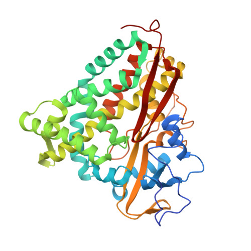
Crystal structures of metyrapone- and phenylimidazole-inhibited complexes of cytochrome P-450cam.

Poulos, T.L.Howard, A.J.

(1987) Biochemistry 26: 8165-8174

  • DOI: https://doi.org/10.1021/bi00399a022
  • Primary Citation of Related Structures:  
    1PHD, 1PHE, 1PHF, 1PHG

  • PubMed Abstract: 

    The crystal structures of metyrapone- and 1-, 2-, and 4-phenylimidazole-inhibited complexes of cytochrome P-450cam have been refined to a nominal resolution of 2.1 A and compared with the 1.63-A camphor-bound structure. With the exception of 2-phenylimidazole, each of the inhibitors forms an N-Fe bond with the heme iron atom while part of the inhibitor sits in the camphor-binding pocket. In the 2-phenylimidazole complex, a water molecule or hydroxide ion coordinates with the heme iron atom while the inhibitor binds in the camphor pocket adjacent to the aqua ligand. Each of the inhibitors forces the central region of helix I that forms part of the O2 binding pocket to move away from the inhibitor, with the exception of 2-phenylimidazole where the helix moves in toward the inhibitor. In addition, the Tyr-96 region, which provides specific contact points with the substrate, is perturbed, although to varying degrees with each inhibitor. These perturbations include large, localized changes in Debye-Waller or temperature factors, indicative of changes in dynamical fluctuations. The largest inhibitor, metyrapone, causes the fewest changes, while 2-phenylimidazole binding causes the largest, especially in helix I. The large 2-phenylimidazole-induced movement of helix I can be rationalized on the basis of the inhibitor imidazole group's hydrogen-bonding requirements.


  • Organizational Affiliation

    Protein Engineering Department, Genex Corporation, Gaithersburg, Maryland 20877.


Macromolecules
Find similar proteins by:  (by identity cutoff)  |  3D Structure
Entity ID: 1
MoleculeChains Sequence LengthOrganismDetailsImage
CYTOCHROME P450-CAM414Pseudomonas putidaMutation(s): 0 
EC: 1.14.15.1
UniProt
Find proteins for P00183 (Pseudomonas putida)
Explore P00183 
Go to UniProtKB:  P00183
Entity Groups  
Sequence Clusters30% Identity50% Identity70% Identity90% Identity95% Identity100% Identity
UniProt GroupP00183
Sequence Annotations
Expand
  • Reference Sequence
Small Molecules
Ligands 2 Unique
IDChains Name / Formula / InChI Key2D Diagram3D Interactions
HEM
Query on HEM

Download Ideal Coordinates CCD File 
B [auth A]PROTOPORPHYRIN IX CONTAINING FE
C34 H32 Fe N4 O4
KABFMIBPWCXCRK-RGGAHWMASA-L
PIW
Query on PIW

Download Ideal Coordinates CCD File 
C [auth A]1-phenyl-1H-imidazole
C9 H8 N2
SEULWJSKCVACTH-UHFFFAOYSA-N
Experimental Data & Validation

Experimental Data

  • Method: X-RAY DIFFRACTION
  • Resolution: 1.60 Å
  • R-Value Observed: 0.190 
  • Space Group: P 21 21 21
Unit Cell:
Length ( Å )Angle ( ˚ )
a = 108.67α = 90
b = 103.9β = 90
c = 36.38γ = 90
Software Package:
Software NamePurpose
PROLSQrefinement

Structure Validation

View Full Validation Report


Entry History 

Deposition Data

  • Released Date: 1993-10-31 
  • Deposition Author(s): Poulos, T.L.

Revision History  (Full details and data files)

  • Version 1.0: 1993-10-31
    Type: Initial release
  • Version 1.1: 2008-03-03
    Changes: Version format compliance
  • Version 1.2: 2011-07-13
    Changes: Non-polymer description, Version format compliance
  • Version 1.3: 2011-07-27
    Changes: Version format compliance
  • Version 1.4: 2017-11-29
    Changes: Derived calculations, Other
  • Version 1.5: 2024-02-14
    Changes: Data collection, Database references, Derived calculations