1P7K

Crystal structure of an anti-ssDNA antigen-binding fragment (Fab) bound to 4-(2-Hydroxyethyl)piperazine-1-ethanesulfonic acid (HEPES)


Experimental Data Snapshot

  • Method: X-RAY DIFFRACTION
  • Resolution: 1.75 Å
  • R-Value Free: 0.230 
  • R-Value Work: 0.197 
  • R-Value Observed: 0.197 

wwPDB Validation   3D Report Full Report


This is version 1.3 of the entry. See complete history


Literature

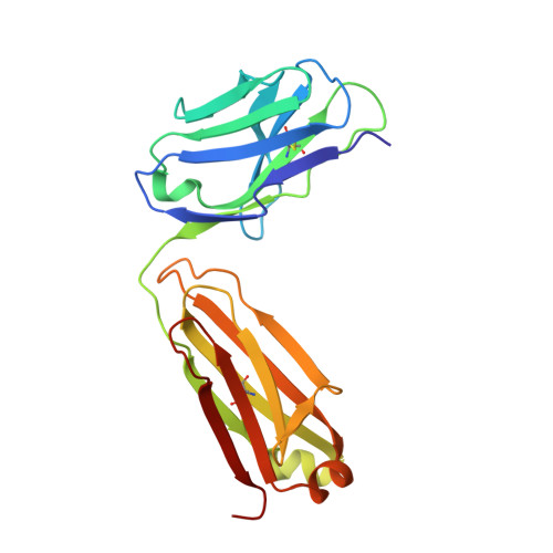
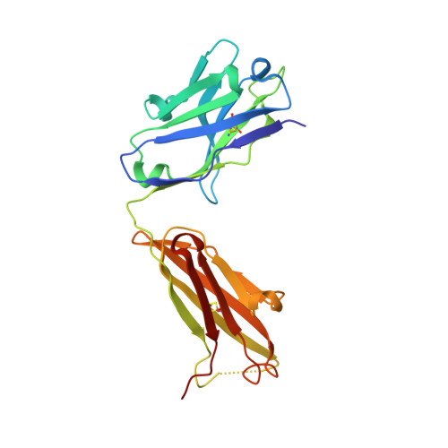
Structure of an anti-DNA fab complexed with a non-DNA ligand provides insights into cross-reactivity and molecular mimicry.

Schuermann, J.P.Henzl, M.T.Deutscher, S.L.Tanner, J.J.

(2004) Proteins 57: 269-278

  • DOI: https://doi.org/10.1002/prot.20200
  • Primary Citation of Related Structures:  
    1P7K

  • PubMed Abstract: 

    Antibodies that recognize DNA (anti-DNA) are part of the autoimmune response underlying systemic lupus erythematosus. To better understand molecular recognition by anti-DNA antibodies, crystallographic studies have been performed using an anti-ssDNA antigen-binding fragment (Fab) known as DNA-1. The previously determined structure of a DNA-1/dT5 complex revealed that thymine bases insert into a narrow groove, and that ligand recognition primarily involves the bases of DNA. We now report the 1.75-A resolution structure of DNA-1 complexed with the biological buffer HEPES (4-(2-Hydroxyethyl)piperazine-1-ethanesulfonic acid). All three light chain complementarity-determining regions (CDRs) and HCDR3 contribute to binding. The HEPES sulfonate hydrogen bonds to His L91, Asn L50, and to the backbone of Tyr H100 and Tyr H100A. The Tyr side-chains of L32, L92, H100, and H100A form nonpolar contacts with the HEPES ethylene and piperazine groups. Comparison to the DNA-1/dT5 structure reveals that the dual recognition of dT5 and HEPES requires a 13-A movement of HCDR3. This dramatic structural change converts the combining site from a narrow groove, appropriate for the edge-on insertion of thymine bases, to one sufficiently wide to accommodate the HEPES sulfonate and piperazine. Isothermal titration calorimetry verified the association of HEPES with DNA-1 under conditions similar those used for crystallization (2 M ammonium sulfate). Interestingly, the presence of 2 M ammonium sulfate increases the affinities of DNA-1 for both HEPES and dT5, suggesting that non-polar Fab-ligand interactions are important for molecular recognition in highly ionic solvent conditions. The structural and thermodynamic data suggest a molecular mimicry mechanism based on structural plasticity and hydrophobic interactions.


  • Organizational Affiliation

    Department of Chemistry, University of Missouri-Columbia, Columbia, Missouri 65211, USA.


Macromolecules
Find similar proteins by:  (by identity cutoff)  |  3D Structure
Entity ID: 1
MoleculeChains Sequence LengthOrganismDetailsImage
antibody light chain FABA [auth L],
C [auth A]
214Mus musculusMutation(s): 0 
UniProt
Find proteins for I6L978 (Mus musculus)
Explore I6L978 
Go to UniProtKB:  I6L978
Entity Groups  
Sequence Clusters30% Identity50% Identity70% Identity90% Identity95% Identity100% Identity
UniProt GroupI6L978
Sequence Annotations
Expand
  • Reference Sequence
Find similar proteins by:  (by identity cutoff)  |  3D Structure
Entity ID: 2
MoleculeChains Sequence LengthOrganismDetailsImage
antibody heavy chain FABB [auth H],
D [auth B]
224Mus musculusMutation(s): 0 
UniProt
Find proteins for P01868 (Mus musculus)
Explore P01868 
Go to UniProtKB:  P01868
Entity Groups  
Sequence Clusters30% Identity50% Identity70% Identity90% Identity95% Identity100% Identity
UniProt GroupP01868
Sequence Annotations
Expand
  • Reference Sequence
Small Molecules
Ligands 5 Unique
IDChains Name / Formula / InChI Key2D Diagram3D Interactions
EPE
Query on EPE

Download Ideal Coordinates CCD File 
EA [auth B],
F [auth L]
4-(2-HYDROXYETHYL)-1-PIPERAZINE ETHANESULFONIC ACID
C8 H18 N2 O4 S
JKMHFZQWWAIEOD-UHFFFAOYSA-N
1PE
Query on 1PE

Download Ideal Coordinates CCD File 
FA [auth B],
G [auth L],
GA [auth B],
S [auth H]
PENTAETHYLENE GLYCOL
C10 H22 O6
JLFNLZLINWHATN-UHFFFAOYSA-N
PEG
Query on PEG

Download Ideal Coordinates CCD File 
H [auth L],
T [auth H]
DI(HYDROXYETHYL)ETHER
C4 H10 O3
MTHSVFCYNBDYFN-UHFFFAOYSA-N
SO4
Query on SO4

Download Ideal Coordinates CCD File 
CA [auth B]
DA [auth B]
E [auth L]
N [auth H]
O [auth H]
CA [auth B],
DA [auth B],
E [auth L],
N [auth H],
O [auth H],
P [auth H],
Q [auth H],
R [auth H],
X [auth A]
SULFATE ION
O4 S
QAOWNCQODCNURD-UHFFFAOYSA-L
GOL
Query on GOL

Download Ideal Coordinates CCD File 
AA [auth A]
BA [auth A]
HA [auth B]
I [auth L]
IA [auth B]
AA [auth A],
BA [auth A],
HA [auth B],
I [auth L],
IA [auth B],
J [auth L],
JA [auth B],
K [auth L],
KA [auth B],
L,
M [auth L],
U [auth H],
V [auth H],
W [auth H],
Y [auth A],
Z [auth A]
GLYCEROL
C3 H8 O3
PEDCQBHIVMGVHV-UHFFFAOYSA-N
Experimental Data & Validation

Experimental Data

  • Method: X-RAY DIFFRACTION
  • Resolution: 1.75 Å
  • R-Value Free: 0.230 
  • R-Value Work: 0.197 
  • R-Value Observed: 0.197 
  • Space Group: P 21 21 2
Unit Cell:
Length ( Å )Angle ( ˚ )
a = 107.666α = 90
b = 156.703β = 90
c = 61.003γ = 90
Software Package:
Software NamePurpose
CNSrefinement
HKL-2000data reduction
SCALEPACKdata scaling
AMoREphasing

Structure Validation

View Full Validation Report


Entry History 

Deposition Data

Revision History  (Full details and data files)

  • Version 1.0: 2004-05-11
    Type: Initial release
  • Version 1.1: 2008-04-29
    Changes: Version format compliance
  • Version 1.2: 2011-07-13
    Changes: Non-polymer description, Version format compliance
  • Version 1.3: 2023-08-16
    Changes: Data collection, Database references, Derived calculations, Refinement description