1ONW

Crystal structure of Isoaspartyl Dipeptidase from E. coli


Experimental Data Snapshot

  • Method: X-RAY DIFFRACTION
  • Resolution: 1.65 Å
  • R-Value Free: 0.219 
  • R-Value Work: 0.178 
  • R-Value Observed: 0.182 

wwPDB Validation   3D Report Full Report


This is version 1.4 of the entry. See complete history


Literature

High Resolution X-ray Structure of Isoaspartyl Dipeptidase from Escherichia coli

Thoden, J.B.Marti-Arbona, R.Raushel, F.M.Holden, H.M.

(2003) Biochemistry 42: 4874-4882

  • DOI: https://doi.org/10.1021/bi034233p
  • Primary Citation of Related Structures:  
    1ONW, 1ONX

  • PubMed Abstract: 

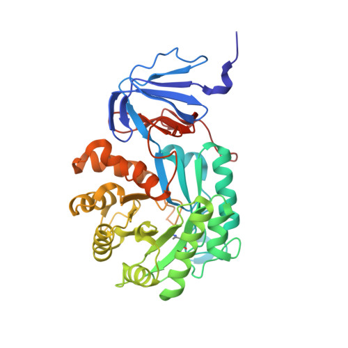
    Isoaspartyl dipeptidase from Escherichia coli functions in protein degradation by catalyzing the hydrolysis of beta-L-isoaspartyl linkages in dipeptides. The best substrate for the enzyme reported thus far is iso-Asp-Leu. Here we report the X-ray analysis of the enzyme in its resting state and complexed with aspartate to 1.65 and 2.1 A resolution, respectively. The quaternary structure of the enzyme is octameric and can be aptly described as a tetramer of dimers. Each subunit folds into two distinct domains: the N-terminal region containing eight strands of mixed beta-sheet and the C-terminal motif that is dominated by a (beta,alpha)(8)-barrel. A binuclear zinc center is located in each subunit at the C-terminal end of the (beta,alpha)(8)-barrel. Ligands to the binuclear metal center include His 68, His 70, His 201, His 230, and Asp 285. The two zincs are bridged by a carboxylated lysine residue (Lys 162) and a solvent molecule, most likely a hydroxide ion. The product of the reaction, aspartate, binds to the enzyme by displacing the bridging solvent with its side chain functional group. From this investigation it is proposed that the reaction mechanism of the enzyme proceeds through a tetrahedral intermediate and that the bridging solvent attacks the re face of the carbonyl carbon of the scissile peptide bond. This structural analysis confirms the placement of isoaspartyl dipeptidase into the urease-related amidohydrolase superfamily.


  • Organizational Affiliation

    Department of Biochemistry, University of Wisconsin, Madison, Wisconsin 53706, USA.


Macromolecules
Find similar proteins by:  (by identity cutoff)  |  3D Structure
Entity ID: 1
MoleculeChains Sequence LengthOrganismDetailsImage
Isoaspartyl dipeptidase
A, B
390Escherichia coliMutation(s): 1 
Gene Names: IADA OR B4328
EC: 3.4.19
UniProt
Find proteins for P39377 (Escherichia coli (strain K12))
Explore P39377 
Go to UniProtKB:  P39377
Entity Groups  
Sequence Clusters30% Identity50% Identity70% Identity90% Identity95% Identity100% Identity
UniProt GroupP39377
Sequence Annotations
Expand
  • Reference Sequence
Small Molecules
Ligands 5 Unique
IDChains Name / Formula / InChI Key2D Diagram3D Interactions
ZN
Query on ZN

Download Ideal Coordinates CCD File 
C [auth A],
D [auth A],
I [auth B],
J [auth B]
ZINC ION
Zn
PTFCDOFLOPIGGS-UHFFFAOYSA-N
EDO
Query on EDO

Download Ideal Coordinates CCD File 
G [auth A],
H [auth A],
N [auth B]
1,2-ETHANEDIOL
C2 H6 O2
LYCAIKOWRPUZTN-UHFFFAOYSA-N
CL
Query on CL

Download Ideal Coordinates CCD File 
E [auth A],
F [auth A],
M [auth B]
CHLORIDE ION
Cl
VEXZGXHMUGYJMC-UHFFFAOYSA-M
MG
Query on MG

Download Ideal Coordinates CCD File 
K [auth B]MAGNESIUM ION
Mg
JLVVSXFLKOJNIY-UHFFFAOYSA-N
NA
Query on NA

Download Ideal Coordinates CCD File 
L [auth B]SODIUM ION
Na
FKNQFGJONOIPTF-UHFFFAOYSA-N
Modified Residues  1 Unique
IDChains TypeFormula2D DiagramParent
KCX
Query on KCX
A, B
L-PEPTIDE LINKINGC7 H14 N2 O4LYS
Experimental Data & Validation

Experimental Data

  • Method: X-RAY DIFFRACTION
  • Resolution: 1.65 Å
  • R-Value Free: 0.219 
  • R-Value Work: 0.178 
  • R-Value Observed: 0.182 
  • Space Group: P 4 21 2
Unit Cell:
Length ( Å )Angle ( ˚ )
a = 116.7α = 90
b = 116.7β = 90
c = 138.5γ = 90
Software Package:
Software NamePurpose
FRAMBOdata collection
SAINTdata reduction
SOLVEphasing
TNTrefinement
SAINTdata scaling

Structure Validation

View Full Validation Report


Entry History 

Deposition Data

Revision History  (Full details and data files)

  • Version 1.0: 2003-05-06
    Type: Initial release
  • Version 1.1: 2008-04-29
    Changes: Version format compliance
  • Version 1.2: 2011-07-13
    Changes: Derived calculations, Version format compliance
  • Version 1.3: 2011-11-16
    Changes: Atomic model
  • Version 1.4: 2017-10-11
    Changes: Refinement description