1OL7

Structure of Human Aurora-A 122-403 phosphorylated on Thr287, Thr288


Experimental Data Snapshot

  • Method: X-RAY DIFFRACTION
  • Resolution: 2.75 Å
  • R-Value Free: 0.296 
  • R-Value Work: 0.257 
  • R-Value Observed: 0.257 

wwPDB Validation   3D Report Full Report


Ligand Structure Quality Assessment 


This is version 1.2 of the entry. See complete history


Literature

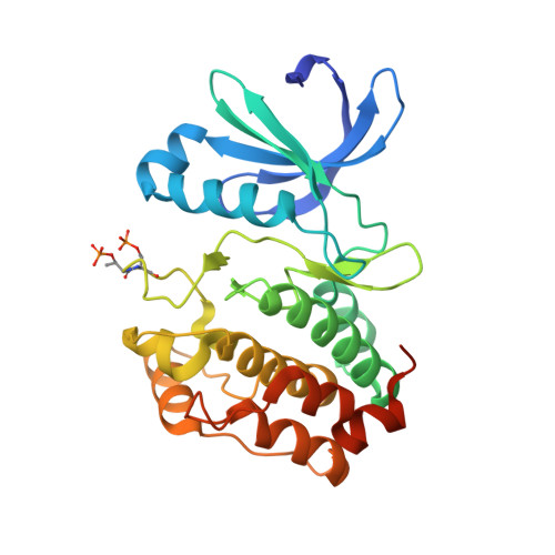
Structural Basis of Aurora-A Activation by Tpx2 at the Mitotic Spindle

Bayliss, R.Sardon, T.Vernos, I.Conti, E.

(2003) Mol Cell 12: 851

  • DOI: https://doi.org/10.1016/s1097-2765(03)00392-7
  • Primary Citation of Related Structures:  
    1OL5, 1OL6, 1OL7

  • PubMed Abstract: 

    Aurora-A is an oncogenic kinase essential for mitotic spindle assembly. It is activated by phosphorylation and by the microtubule-associated protein TPX2, which also localizes the kinase to spindle microtubules. We have uncovered the molecular mechanism of Aurora-A activation by determining crystal structures of its phosphorylated form both with and without a 43 residue long domain of TPX2 that we identified as fully functional for kinase activation and protection from dephosphorylation. In the absence of TPX2, the Aurora-A activation segment is in an inactive conformation, with the crucial phosphothreonine exposed and accessible for deactivation. Binding of TPX2 triggers no global conformational changes in the kinase but pulls on the activation segment, swinging the phosphothreonine into a buried position and locking the active conformation. The recognition between Aurora-A and TPX2 resembles that between the cAPK catalytic core and its flanking regions, suggesting this molecular mechanism may be a recurring theme in kinase regulation.


  • Organizational Affiliation

    Structural and Computational Biology Programme, European Molecular Biology Laboratory, Meyerhofstrasse 1, D-69117, Heidelberg, Germany .


Macromolecules
Find similar proteins by:  (by identity cutoff)  |  3D Structure
Entity ID: 1
MoleculeChains Sequence LengthOrganismDetailsImage
SERINE/THREONINE KINASE 6282Homo sapiensMutation(s): 0 
EC: 2.7.1.37
UniProt & NIH Common Fund Data Resources
Find proteins for O14965 (Homo sapiens)
Explore O14965 
Go to UniProtKB:  O14965
PHAROS:  O14965
GTEx:  ENSG00000087586 
Entity Groups  
Sequence Clusters30% Identity50% Identity70% Identity90% Identity95% Identity100% Identity
UniProt GroupO14965
Sequence Annotations
Expand
  • Reference Sequence
Small Molecules
Modified Residues  1 Unique
IDChains TypeFormula2D DiagramParent
TPO
Query on TPO
A
L-PEPTIDE LINKINGC4 H10 N O6 PTHR
Experimental Data & Validation

Experimental Data

  • Method: X-RAY DIFFRACTION
  • Resolution: 2.75 Å
  • R-Value Free: 0.296 
  • R-Value Work: 0.257 
  • R-Value Observed: 0.257 
  • Space Group: P 61 2 2
Unit Cell:
Length ( Å )Angle ( ˚ )
a = 81.18α = 90
b = 81.18β = 90
c = 169.62γ = 120
Software Package:
Software NamePurpose
CNSrefinement
MOSFLMdata reduction
SCALAdata scaling
CNSphasing

Structure Validation

View Full Validation Report


Ligand Structure Quality Assessment 


Entry History 

Deposition Data

Revision History  (Full details and data files)

  • Version 1.0: 2003-10-30
    Type: Initial release
  • Version 1.1: 2011-05-08
    Changes: Version format compliance
  • Version 1.2: 2011-07-13
    Changes: Version format compliance