1OBY

Crystal structure of the complex of PDZ2 of syntenin with a syndecan-4 peptide.


Experimental Data Snapshot

  • Method: X-RAY DIFFRACTION
  • Resolution: 1.85 Å
  • R-Value Free: 0.226 
  • R-Value Work: 0.175 

wwPDB Validation   3D Report Full Report


This is version 1.5 of the entry. See complete history


Literature

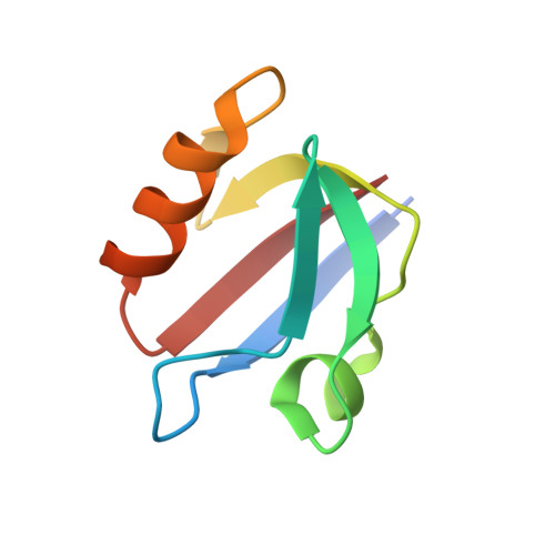
Molecular Roots of Degenerate Specificity in Syntenin'S Pdz2 Domain: Reassessment of the Pdz Recognition Paradigm

Kang, B.S.Cooper, D.R.Devedjiev, Y.Derewenda, U.Derewenda, Z.S.

(2003) Structure 11: 845

  • DOI: https://doi.org/10.1016/s0969-2126(03)00125-4
  • Primary Citation of Related Structures:  
    1NTE, 1OBX, 1OBY, 1OBZ

  • PubMed Abstract: 

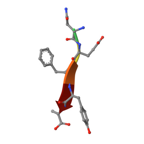
    Crystal structures of the PDZ2 domain of the scaffolding protein syntenin, both unbound and in complexes with peptides derived from C termini of IL5 receptor (alpha chain) and syndecan, reveal the molecular roots of syntenin's degenerate specificity. Three distinct binding sites (S(0), S(-1), and S(-2)), with affinities for hydrophobic side chains, function in a combinatorial way: S(-1) and S(-2) act together to bind syndecan, while S(0) and S(-1) are involved in the binding of IL5Ralpha. Neither mode of interaction is consistent with the prior classification scheme, which defined the IL5Ralpha interaction as class I (-S/T-X-phi) and the syndecan interaction as class II (-phi-X-phi). These results, in conjunction with other emerging structural data on PDZ domains, call for a revision of their classification and of the existing model of their mechanism.


  • Organizational Affiliation

    Department of Molecular Physiology and Biological Physics, The Cancer Center, University of Virginia, Charlottesville, VA 22908, USA.


Macromolecules
Find similar proteins by:  (by identity cutoff)  |  3D Structure
Entity ID: 1
MoleculeChains Sequence LengthOrganismDetailsImage
SYNTENIN 1
A, B
79Homo sapiensMutation(s): 0 
UniProt & NIH Common Fund Data Resources
Find proteins for O00560 (Homo sapiens)
Explore O00560 
Go to UniProtKB:  O00560
PHAROS:  O00560
GTEx:  ENSG00000137575 
Entity Groups  
Sequence Clusters30% Identity50% Identity70% Identity90% Identity95% Identity100% Identity
UniProt GroupO00560
Sequence Annotations
Expand
  • Reference Sequence

Find similar proteins by:  Sequence   |   3D Structure  

Entity ID: 2
MoleculeChains Sequence LengthOrganismDetailsImage
SYNDECAN-4C [auth P],
D [auth Q]
6Homo sapiensMutation(s): 0 
UniProt & NIH Common Fund Data Resources
Find proteins for P31431 (Homo sapiens)
Explore P31431 
Go to UniProtKB:  P31431
PHAROS:  P31431
GTEx:  ENSG00000124145 
Entity Groups  
UniProt GroupP31431
Sequence Annotations
Expand
  • Reference Sequence
Experimental Data & Validation

Experimental Data

  • Method: X-RAY DIFFRACTION
  • Resolution: 1.85 Å
  • R-Value Free: 0.226 
  • R-Value Work: 0.175 
  • Space Group: C 1 2 1
Unit Cell:
Length ( Å )Angle ( ˚ )
a = 58.337α = 90
b = 54.441β = 98.65
c = 50.219γ = 90
Software Package:
Software NamePurpose
REFMACrefinement
HKL-2000data reduction
HKL-2000data scaling
AMoREphasing

Structure Validation

View Full Validation Report


Entry History 

Deposition Data

Revision History  (Full details and data files)

  • Version 1.0: 2003-07-11
    Type: Initial release
  • Version 1.1: 2011-05-08
    Changes: Version format compliance
  • Version 1.2: 2011-07-13
    Changes: Version format compliance
  • Version 1.3: 2019-05-08
    Changes: Data collection, Experimental preparation, Other
  • Version 1.4: 2019-10-16
    Changes: Data collection, Other
  • Version 1.5: 2023-12-13
    Changes: Data collection, Database references, Refinement description