1NY3

Crystal structure of ADP bound to MAP KAP kinase 2


Experimental Data Snapshot

  • Method: X-RAY DIFFRACTION
  • Resolution: 3.00 Å
  • R-Value Free: 0.292 
  • R-Value Work: 0.259 
  • R-Value Observed: 0.259 

wwPDB Validation   3D Report Full Report


This is version 1.3 of the entry. See complete history


Literature

Catalytically active MAP KAP kinase 2 structures in complex with staurosporine and ADP reveal differences with the autoinhibited enzyme

Underwood, K.W.Parris, K.D.Federico, E.Mosyak, L.Czerwinski, R.M.Shane, T.Taylor, M.Svenson, K.Liu, Y.Hsiao, C.L.Wolfrom, S.Maguire, M.Malakian, K.Telliez, J.B.Lin, L.L.Kriz, R.W.Seehra, J.Somers, W.S.Stahl, M.L.

(2003) Structure 11: 627-636

  • DOI: https://doi.org/10.1016/s0969-2126(03)00092-3
  • Primary Citation of Related Structures:  
    1NXK, 1NY3

  • PubMed Abstract: 

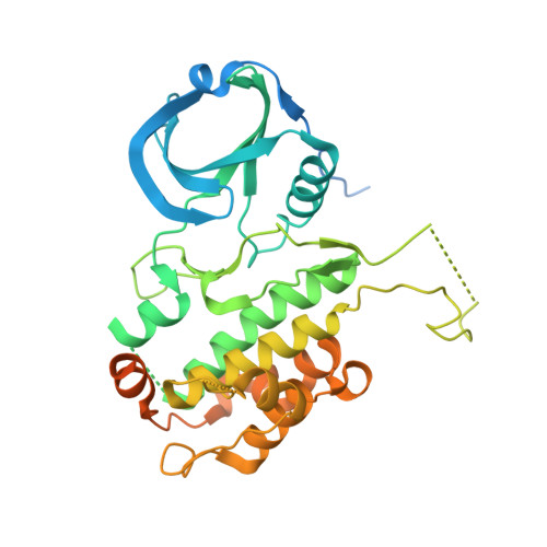
    MAP KAP kinase 2 (MK2), a Ser/Thr kinase, plays a crucial role in the inflammatory process. We have determined the crystal structures of a catalytically active C-terminal deletion form of human MK2, residues 41-364, in complex with staurosporine at 2.7 A and with ADP at 3.2 A, revealing overall structural similarity with other Ser/Thr kinases. Kinetic analysis reveals that the K(m) for ATP is very similar for MK2 41-364 and p38-activated MK2 41-400. Conversely, the catalytic rate and binding for peptide substrate are dramatically reduced in MK2 41-364. However, phosphorylation of MK2 41-364 by p38 restores the V(max) and K(m) for peptide substrate to values comparable to those seen in p38-activated MK2 41-400, suggesting a mechanism for regulation of enzyme activity.


  • Organizational Affiliation

    Department of Biological Chemistry, Wyeth Research, 87 Cambridge Park Drive, Cambridge, MA 02140, USA. kunderwood@wyeth.com


Macromolecules
Find similar proteins by:  (by identity cutoff)  |  3D Structure
Entity ID: 1
MoleculeChains Sequence LengthOrganismDetailsImage
MAP kinase-activated protein kinase 2400Homo sapiensMutation(s): 0 
Gene Names: MAPKAPK2
EC: 2.7.1
UniProt & NIH Common Fund Data Resources
Find proteins for P49137 (Homo sapiens)
Explore P49137 
Go to UniProtKB:  P49137
PHAROS:  P49137
GTEx:  ENSG00000162889 
Entity Groups  
Sequence Clusters30% Identity50% Identity70% Identity90% Identity95% Identity100% Identity
UniProt GroupP49137
Sequence Annotations
Expand
  • Reference Sequence
Small Molecules
Ligands 1 Unique
IDChains Name / Formula / InChI Key2D Diagram3D Interactions
ADP
Query on ADP

Download Ideal Coordinates CCD File 
B [auth A]ADENOSINE-5'-DIPHOSPHATE
C10 H15 N5 O10 P2
XTWYTFMLZFPYCI-KQYNXXCUSA-N
Experimental Data & Validation

Experimental Data

  • Method: X-RAY DIFFRACTION
  • Resolution: 3.00 Å
  • R-Value Free: 0.292 
  • R-Value Work: 0.259 
  • R-Value Observed: 0.259 
  • Space Group: F 41 3 2
Unit Cell:
Length ( Å )Angle ( ˚ )
a = 253.049α = 90
b = 253.049β = 90
c = 253.049γ = 90
Software Package:
Software NamePurpose
REFMACrefinement
SCALEPACKdata scaling
AMoREphasing

Structure Validation

View Full Validation Report


Entry History 

Revision History  (Full details and data files)

  • Version 1.0: 2003-10-14
    Type: Initial release
  • Version 1.1: 2008-04-29
    Changes: Version format compliance
  • Version 1.2: 2011-07-13
    Changes: Derived calculations, Version format compliance
  • Version 1.3: 2023-08-16
    Changes: Data collection, Database references, Derived calculations, Refinement description