1NXC

Structure of mouse Golgi alpha-1,2-mannosidase IA reveals the molecular basis for substrate specificity among Class I enzymes (family 47 glycosidases)


Experimental Data Snapshot

  • Method: X-RAY DIFFRACTION
  • Resolution: 1.51 Å
  • R-Value Free: 0.190 
  • R-Value Work: 0.171 
  • R-Value Observed: 0.172 

wwPDB Validation   3D Report Full Report


This is version 2.0 of the entry. See complete history


Literature

Structure of mouse Golgi alpha-mannosidase IA reveals the molecular basis for substrate specificity among class 1 (family 47 glycosylhydrolase) alpha1,2-mannosidases

Tempel, W.Karaveg, K.Liu, Z.-J.Rose, J.Wang, B.-C.Moremen, K.W.

(2004) J Biol Chem 279: 29774-29786

  • DOI: https://doi.org/10.1074/jbc.M403065200
  • Primary Citation of Related Structures:  
    1NXC

  • PubMed Abstract: 

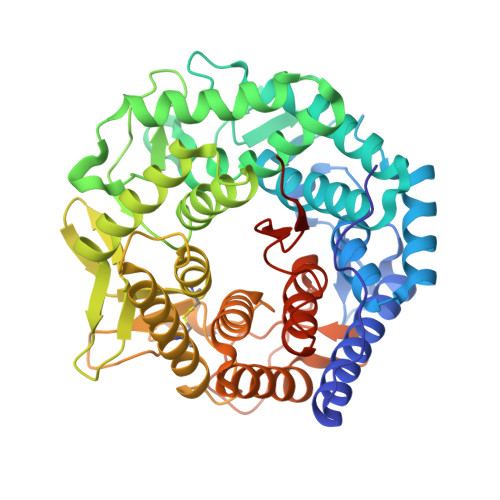
    Three subfamilies of mammalian Class 1 processing alpha1,2-mannosidases (family 47 glycosidases) play critical roles in the maturation of Asn-linked glycoproteins in the endoplasmic reticulum (ER) and Golgi complex as well as influencing the timing and recognition for disposal of terminally unfolded proteins by ER-associated degradation. In an effort to define the structural basis for substrate recognition among Class 1 mannosidases, we have crystallized murine Golgi mannosidase IA (space group P2(1)2(1)2(1)), and the structure was solved to 1.5-A resolution by molecular replacement. The enzyme assumes an (alphaalpha)(7) barrel structure with a Ca(2+) ion coordinated at the base of the barrel similar to other Class 1 mannosidases. Critical residues within the barrel structure that coordinate the Ca(2+) ion or presumably bind and catalyze the hydrolysis of the glycone are also highly conserved. A Man(6)GlcNAc(2) oligosaccharide attached to Asn(515) in the murine enzyme was found to extend into the active site of an adjoining protein unit in the crystal lattice in a presumed enzyme-product complex. In contrast to an analogous complex previously isolated for Saccharomyces cerevisiae ER mannosidase I, the oligosaccharide in the active site of the murine Golgi enzyme assumes a different conformation to present an alternate oligosaccharide branch into the active site pocket. A comparison of the observed protein-carbohydrate interactions for the murine Golgi enzyme with the binding cleft topologies of the other family 47 glycosidases provides a framework for understanding the structural basis for substrate recognition among this class of enzymes.


  • Organizational Affiliation

    Department of Biochemistry and Molecular Biology, University of Georgia, Athens, Georgia 30602, USA.


Macromolecules
Find similar proteins by:  (by identity cutoff)  |  3D Structure
Entity ID: 1
MoleculeChains Sequence LengthOrganismDetailsImage
Mannosyl-oligosaccharide 1,2-alpha-mannosidase IA478Mus musculusMutation(s): 0 
Gene Names: man1a
EC: 3.2.1.113
UniProt
Find proteins for P45700 (Mus musculus)
Explore P45700 
Go to UniProtKB:  P45700
Entity Groups  
Sequence Clusters30% Identity50% Identity70% Identity90% Identity95% Identity100% Identity
UniProt GroupP45700
Sequence Annotations
Expand
  • Reference Sequence
Oligosaccharides

Help

Entity ID: 2
MoleculeChains Length2D Diagram Glycosylation3D Interactions
alpha-D-mannopyranose-(1-6)-alpha-D-mannopyranose-(1-3)-[alpha-D-mannopyranose-(1-3)-[alpha-D-mannopyranose-(1-6)]alpha-D-mannopyranose-(1-6)]beta-D-mannopyranose-(1-4)-2-acetamido-2-deoxy-beta-D-glucopyranose-(1-4)-2-acetamido-2-deoxy-beta-D-glucopyranose
B
8N-Glycosylation
Glycosylation Resources
GlyTouCan:  G92684AL
GlyCosmos:  G92684AL
GlyGen:  G92684AL
Small Molecules
Ligands 1 Unique
IDChains Name / Formula / InChI Key2D Diagram3D Interactions
CA
Query on CA

Download Ideal Coordinates CCD File 
C [auth A]CALCIUM ION
Ca
BHPQYMZQTOCNFJ-UHFFFAOYSA-N
Experimental Data & Validation

Experimental Data

  • Method: X-RAY DIFFRACTION
  • Resolution: 1.51 Å
  • R-Value Free: 0.190 
  • R-Value Work: 0.171 
  • R-Value Observed: 0.172 
  • Space Group: P 21 21 21
Unit Cell:
Length ( Å )Angle ( ˚ )
a = 55.288α = 90
b = 72.164β = 90
c = 129.571γ = 90
Software Package:
Software NamePurpose
EPMRphasing
REFMACrefinement
PROTEUM PLUSdata reduction

Structure Validation

View Full Validation Report


Entry History 

Revision History  (Full details and data files)

  • Version 1.0: 2004-05-04
    Type: Initial release
  • Version 1.1: 2008-04-29
    Changes: Version format compliance
  • Version 1.2: 2011-07-13
    Changes: Non-polymer description, Version format compliance
  • Version 1.3: 2017-10-11
    Changes: Refinement description
  • Version 2.0: 2020-07-29
    Type: Remediation
    Reason: Carbohydrate remediation
    Changes: Atomic model, Data collection, Derived calculations, Structure summary