1NLB

crystal structure of anti-HCV monoclonal antibody 19D9D6


Experimental Data Snapshot

  • Method: X-RAY DIFFRACTION
  • Resolution: 1.60 Å
  • R-Value Free: 0.211 
  • R-Value Work: 0.181 
  • R-Value Observed: 0.197 

wwPDB Validation   3D Report Full Report


This is version 1.4 of the entry. See complete history


Literature

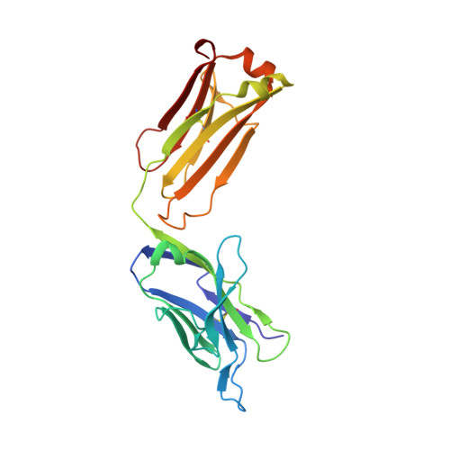
Crystal structure of a hydrophobic immunodominant antigenic site on hepatitis C virus core protein complexed to monoclonal antibody 19D9D6.

Menez, R.Bossus, M.Muller, B.H.Sibai, G.Dalbon, P.Ducancel, F.Jolivet-Reynaud, C.Stura, E.A.

(2003) J Immunol 170: 1917-1924

  • DOI: https://doi.org/10.4049/jimmunol.170.4.1917
  • Primary Citation of Related Structures:  
    1N64, 1NLB

  • PubMed Abstract: 

    The first crystal structure of a complex between a hepatitis C virus (HCV) core protein-derived peptide (residues 13-40) and the Ab fragment of a murine mAb (19D9D6) has been solved, allowing determination of the recognized epitope and elucidation of its conformation. This Ab, raised against the first 120 residues of the core protein, recognizes core particles and strongly competes with anticore human Abs, suggesting that it is highly representative of the human anti-HCV core response. Its epitope lies within the first 45 aa of the protein, the major antigenic segment of core recognized both by murine and human Abs. Surprisingly, the recognized epitope (29-37: QIVGGVYLL) has an unusual preponderance of hydrophobic residues, some of which are buried in a small hydrophobic core in the nuclear magnetic resonance structure of the peptide (2-45) in solution, suggesting that the Ab may induce a structural rearrangement upon recognition. The flexibility may reside entirely within the Ag, since the Fab'-peptide complex structure at 2.34 A shows that the Ab binding site is hardly perturbed by complexation. Given that the recognized residues are unlikely to be solvent exposed, we are left with the interesting possibility that Ab-core interactions may take place in a nonaqueous environment.


  • Organizational Affiliation

    Unité Mixte Commissariat à l'Energie Atomique, bioMérieux and Département d'Ingénierie et d'Etudes des Protéines, Commissariat à l'Energie Atomique, Centre d'Etudes de Saclay, Gif-sur-Yvette, France.


Macromolecules
Find similar proteins by:  (by identity cutoff)  |  3D Structure
Entity ID: 1
MoleculeChains Sequence LengthOrganismDetailsImage
antibody 19D9D6 light chainA [auth L]220Mus musculusMutation(s): 0 
Entity Groups  
Sequence Clusters30% Identity50% Identity70% Identity90% Identity95% Identity100% Identity
Sequence Annotations
Expand
  • Reference Sequence
Find similar proteins by:  (by identity cutoff)  |  3D Structure
Entity ID: 2
MoleculeChains Sequence LengthOrganismDetailsImage
antibody 19D9D6 heavy chainB [auth H]218Mus musculusMutation(s): 0 
Entity Groups  
Sequence Clusters30% Identity50% Identity70% Identity90% Identity95% Identity100% Identity
Sequence Annotations
Expand
  • Reference Sequence
Experimental Data & Validation

Experimental Data

  • Method: X-RAY DIFFRACTION
  • Resolution: 1.60 Å
  • R-Value Free: 0.211 
  • R-Value Work: 0.181 
  • R-Value Observed: 0.197 
  • Space Group: P 21 21 2
Unit Cell:
Length ( Å )Angle ( ˚ )
a = 63.48α = 90
b = 173.68β = 90
c = 41.73γ = 90
Software Package:
Software NamePurpose
DENZOdata reduction
SCALEPACKdata scaling
AMoREphasing
CNSrefinement

Structure Validation

View Full Validation Report


Entry History 

Deposition Data

Revision History  (Full details and data files)

  • Version 1.0: 2003-02-25
    Type: Initial release
  • Version 1.1: 2008-04-29
    Changes: Version format compliance
  • Version 1.2: 2011-07-13
    Changes: Version format compliance
  • Version 1.3: 2013-09-18
    Changes: Source and taxonomy
  • Version 1.4: 2023-08-16
    Changes: Data collection, Database references, Derived calculations, Refinement description