Distal pocket polarity in ligand binding to myoglobin: structural and functional characterization of a threonine68(E11) mutant.
Smerdon, S.J., Dodson, G.G., Wilkinson, A.J., Gibson, Q.H., Blackmore, R.S., Carver, T.E., Olson, J.S.(1991) Biochemistry 30: 6252-6260
- PubMed: 1905570
- DOI: https://doi.org/10.1021/bi00239a025
- Primary Citation of Related Structures:
1MYJ - PubMed Abstract:
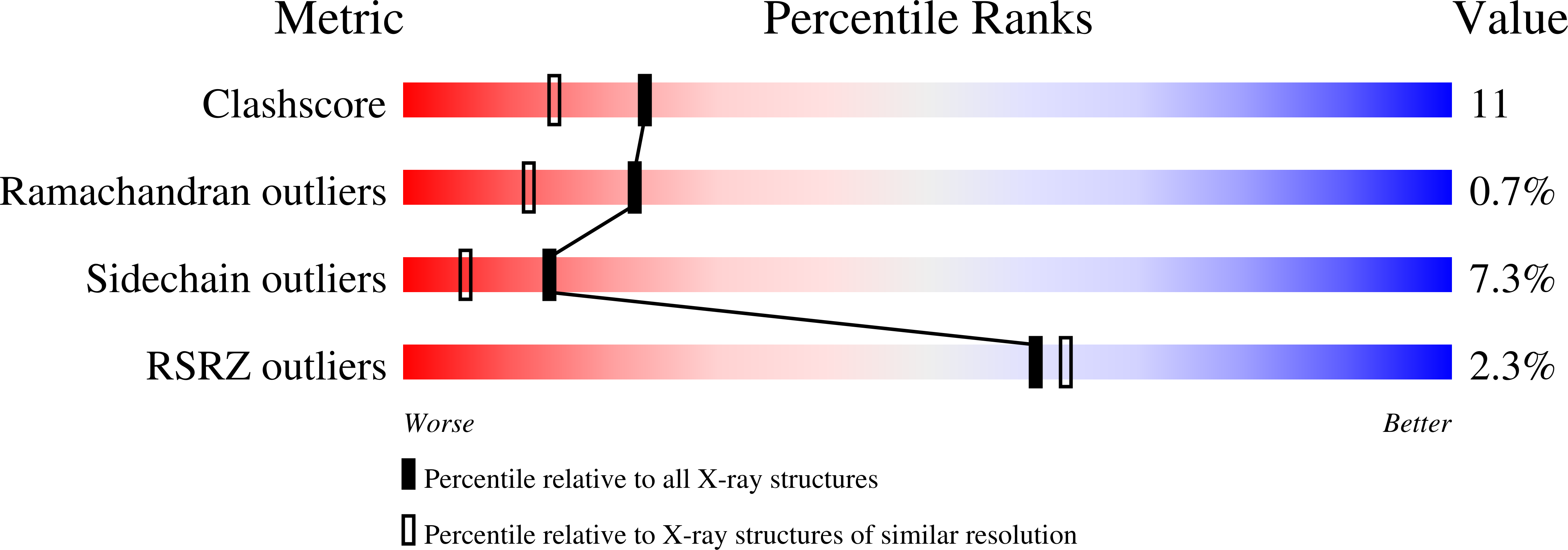
Site-directed mutagenesis studies have confirmed that the distal histidine in myoglobin stabilizes bound O2 by hydrogen bonding and have suggested that it is the polar character of the imidazole side chain rather than its size that limits the rate of ligand entry into the protein. We constructed an isosteric Val68 to Thr replacement in pig myoglobin (i) to investigate whether the O2 affinity could be increased by the introduction of a second hydrogen-bonding group into the distal heme pocket and (ii) to examine the influence of polarity on the ligand binding rates more rigorously. The 1.9-A crystal structure of Thr68 aquometmyoglobin confirms that the mutant and wild-type proteins are essentially isostructural and reveals that the beta-OH group of Thr68 is in a position to form hydrogen-bonding interactions both with the coordinated water molecule and with the main chain greater than C=O of residue 64. The rate of azide binding to the ferric form of the Thr68 mutant was 60-fold lower than that for the wild-type protein, consistent with the proposed stabilization of the coordinated water molecule. However, bound O2 is destabilized in the ferrous form of the mutant protein. The observed 17-fold lowering of the O2 affinity may be a consequence of the hydrogen-bonding interaction made between the Thr68 beta-OH group and the carbonyl oxygen of residue 64. Overall association rate constants for O2, NO, and alkyl isocyanide binding to ferrous pig myoglobin were 3-10-fold lower for the mutant compared to the wild-type protein, whereas that for CO binding was little affected.(ABSTRACT TRUNCATED AT 250 WORDS)
Organizational Affiliation:
Department of Chemistry, University of York, Heslington, U.K.


