1MTW

FACTOR XA SPECIFIC INHIBITOR IN COMPLEX WITH BOVINE TRYPSIN


Experimental Data Snapshot

  • Method: X-RAY DIFFRACTION
  • Resolution: 1.90 Å
  • R-Value Work: 0.169 
  • R-Value Observed: 0.169 

wwPDB Validation   3D Report Full Report


Ligand Structure Quality Assessment 


This is version 1.2 of the entry. See complete history


Literature

Crystal structures of factor Xa specific inhibitors in complex with trypsin: structural grounds for inhibition of factor Xa and selectivity against thrombin.

Stubbs, M.T.Huber, R.Bode, W.

(1995) FEBS Lett 375: 103-107

  • DOI: https://doi.org/10.1016/0014-5793(95)01190-p
  • Primary Citation of Related Structures:  
    1MTS, 1MTU, 1MTV, 1MTW

  • PubMed Abstract: 

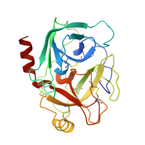
    Crystal structures of DX9065a and a related bisamidino-aryl inhibitor specific for the blood-clotting factor Xa have been solved in complex with bovine beta-trypsin to a resolution of 1.9 A. Each inhibitor exhibits an extended conformation along the active site, in contrast to the compact folded structures observed for thrombin specific inhibitors. Few direct contacts (predominantly in the S1 pocket) are made between trypsin and the inhibitors. Transfer of the inhibitors to the active site of factor Xa suggests a three-site interaction: salt bridge formation at the base of the primary specificity pocket, extensive hydrophobic surface burial and a weak electrostatic interaction between the distal basic component of the inhibitor and an electronegative cavity of factor Xa formed by three backbone carbonyl oxygens. Additivity of these three interactions is the basis for the observed strong inhibition of factor Xa and provides a framework for the design of novel factor Xa inhibitors. A propionic acid group of the inhibitor would clash with the thrombin specific '60-insertion loop', thus conferring selectivity against thrombin.


  • Organizational Affiliation

    Max-Planck Institut für Biochemie, Martinsried bei München, Germany.


Macromolecules
Find similar proteins by:  (by identity cutoff)  |  3D Structure
Entity ID: 1
MoleculeChains Sequence LengthOrganismDetailsImage
TRYPSIN223Bos taurusMutation(s): 0 
EC: 3.4.21.4
UniProt
Find proteins for P00760 (Bos taurus)
Explore P00760 
Go to UniProtKB:  P00760
Entity Groups  
Sequence Clusters30% Identity50% Identity70% Identity90% Identity95% Identity100% Identity
UniProt GroupP00760
Sequence Annotations
Expand
  • Reference Sequence
Small Molecules
Ligands 2 Unique
IDChains Name / Formula / InChI Key2D Diagram3D Interactions
DX9
Query on DX9

Download Ideal Coordinates CCD File 
C [auth A](2S)-3-(7-carbamimidoylnaphthalen-2-yl)-2-[4-({(3R)-1-[(1Z)-ethanimidoyl]pyrrolidin-3-yl}oxy)phenyl]propanoic acid
C26 H28 N4 O3
AGRCGQSFFMCBRE-DGUCVARSSA-N
CA
Query on CA

Download Ideal Coordinates CCD File 
B [auth A]CALCIUM ION
Ca
BHPQYMZQTOCNFJ-UHFFFAOYSA-N
Binding Affinity Annotations 
IDSourceBinding Affinity
DX9 BindingDB:  1MTW Ki: min: 190, max: 200 (nM) from 2 assay(s)
Experimental Data & Validation

Experimental Data

  • Method: X-RAY DIFFRACTION
  • Resolution: 1.90 Å
  • R-Value Work: 0.169 
  • R-Value Observed: 0.169 
  • Space Group: P 21 21 21
Unit Cell:
Length ( Å )Angle ( ˚ )
a = 54.9α = 90
b = 61β = 90
c = 64.3γ = 90
Software Package:
Software NamePurpose
XDSdata scaling
XSCALEdata scaling
X-PLORmodel building
X-PLORrefinement
XDSdata reduction
X-PLORphasing

Structure Validation

View Full Validation Report


Ligand Structure Quality Assessment 


Entry History 

Deposition Data

  • Released Date: 1997-11-12 
  • Deposition Author(s): Stubbs, M.T.

Revision History  (Full details and data files)

  • Version 1.0: 1997-11-12
    Type: Initial release
  • Version 1.1: 2008-03-03
    Changes: Version format compliance
  • Version 1.2: 2011-07-13
    Changes: Version format compliance