1MOQ

ISOMERASE DOMAIN OF GLUCOSAMINE 6-PHOSPHATE SYNTHASE COMPLEXED WITH GLUCOSAMINE 6-PHOSPHATE


Experimental Data Snapshot

  • Method: X-RAY DIFFRACTION
  • Resolution: 1.57 Å
  • R-Value Work: 0.185 

wwPDB Validation   3D Report Full Report


Ligand Structure Quality Assessment 


This is version 1.4 of the entry. See complete history


Literature

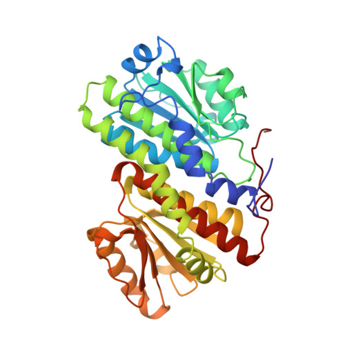
Involvement of the C terminus in intramolecular nitrogen channeling in glucosamine 6-phosphate synthase: evidence from a 1.6 A crystal structure of the isomerase domain.

Teplyakov, A.Obmolova, G.Badet-Denisot, M.A.Badet, B.Polikarpov, I.

(1998) Structure 6: 1047-1055

  • DOI: https://doi.org/10.1016/s0969-2126(98)00105-1
  • Primary Citation of Related Structures:  
    1MOQ

  • PubMed Abstract: 

    Glucosamine 6-phosphate synthase (GlmS) catalyses the first step in hexosamine metabolism, converting fructose-6P (6 phosphate) into glucosamine-6P using glutamine as a nitrogen source. GlmS is a bienzyme complex consisting of two domains that catalyse glutamine hydrolysis and sugar-phosphate isomerisation, respectively. Knowledge of the three-dimensional structure of GlmS is essential for understanding the general principles of catalysis by ketol isomerases and the mechanism of nitrogen transfer in glutamine amidotransferases.


  • Organizational Affiliation

    European Molecular Biology Laboratory, Germany. alexeyt@intra.niddk.nih.gov


Macromolecules
Find similar proteins by:  (by identity cutoff)  |  3D Structure
Entity ID: 1
MoleculeChains Sequence LengthOrganismDetailsImage
GLUCOSAMINE 6-PHOSPHATE SYNTHASE368Escherichia coliMutation(s): 0 
EC: 2.6.1.16
UniProt
Find proteins for P17169 (Escherichia coli (strain K12))
Explore P17169 
Go to UniProtKB:  P17169
Entity Groups  
Sequence Clusters30% Identity50% Identity70% Identity90% Identity95% Identity100% Identity
UniProt GroupP17169
Sequence Annotations
Expand
  • Reference Sequence
Small Molecules
Ligands 5 Unique
IDChains Name / Formula / InChI Key2D Diagram3D Interactions
GLP
Query on GLP

Download Ideal Coordinates CCD File 
B [auth A]2-amino-2-deoxy-6-O-phosphono-alpha-D-glucopyranose
C6 H14 N O8 P
XHMJOUIAFHJHBW-UKFBFLRUSA-N
MES
Query on MES

Download Ideal Coordinates CCD File 
I [auth A]2-(N-MORPHOLINO)-ETHANESULFONIC ACID
C6 H13 N O4 S
SXGZJKUKBWWHRA-UHFFFAOYSA-N
MRD
Query on MRD

Download Ideal Coordinates CCD File 
J [auth A](4R)-2-METHYLPENTANE-2,4-DIOL
C6 H14 O2
SVTBMSDMJJWYQN-RXMQYKEDSA-N
SO4
Query on SO4

Download Ideal Coordinates CCD File 
C [auth A],
D [auth A],
E [auth A],
F [auth A],
G [auth A]
SULFATE ION
O4 S
QAOWNCQODCNURD-UHFFFAOYSA-L
NA
Query on NA

Download Ideal Coordinates CCD File 
H [auth A]SODIUM ION
Na
FKNQFGJONOIPTF-UHFFFAOYSA-N
Binding Affinity Annotations 
IDSourceBinding Affinity
GLP Binding MOAD:  1MOQ Ki: 3.50e+5 (nM) from 1 assay(s)
PDBBind:  1MOQ Ki: 3.50e+5 (nM) from 1 assay(s)
Experimental Data & Validation

Experimental Data

  • Method: X-RAY DIFFRACTION
  • Resolution: 1.57 Å
  • R-Value Work: 0.185 
  • Space Group: H 3 2
Unit Cell:
Length ( Å )Angle ( ˚ )
a = 143.7α = 90
b = 143.7β = 90
c = 173.6γ = 120
Software Package:
Software NamePurpose
CCP4model building
PROLSQrefinement
DENZOdata reduction
SCALEPACKdata scaling
CCP4phasing

Structure Validation

View Full Validation Report


Ligand Structure Quality Assessment 


Entry History 

Deposition Data

Revision History  (Full details and data files)

  • Version 1.0: 1998-10-07
    Type: Initial release
  • Version 1.1: 2008-03-24
    Changes: Version format compliance
  • Version 1.2: 2011-07-13
    Changes: Derived calculations, Version format compliance
  • Version 1.3: 2020-07-29
    Type: Remediation
    Reason: Carbohydrate remediation
    Changes: Data collection, Derived calculations, Other, Structure summary
  • Version 1.4: 2023-08-09
    Changes: Database references, Refinement description, Structure summary