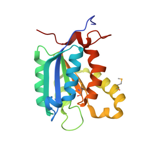
1MKZ

Crystal structure of MoaB protein at 1.6 A resolution.


Experimental Data Snapshot

  • Method: X-RAY DIFFRACTION
  • Resolution: 1.60 Å
  • R-Value Free: 0.219 
  • R-Value Work: 0.183 
  • R-Value Observed: 0.185 

wwPDB Validation   3D Report Full Report


This is version 1.5 of the entry. See complete history


Literature

The crystal structure of Escherichia coli MoaB suggests a probable role in molybdenum cofactor synthesis.

Sanishvili, R.Beasley, S.Skarina, T.Glesne, D.Joachimiak, A.Edwards, A.Savchenko, A.

(2004) J Biol Chem 279: 42139-42146

  • DOI: https://doi.org/10.1074/jbc.M407694200
  • Primary Citation of Related Structures:  
    1MKZ

  • PubMed Abstract: 

    The crystal structure of Escherichia coli MoaB was determined by multiwavelength anomalous diffraction phasing and refined at 1.6-A resolution. The molecule displayed a modified Rossman fold. MoaB is assembled into a hexamer composed of two trimers. The monomers have high structural similarity with two proteins, MogA and MoeA, from the molybdenum cofactor synthesis pathway in E. coli, as well as with domains of mammalian gephyrin and plant Cnx1, which are also involved in molybdopterin synthesis. Structural comparison between these proteins and the amino acid conservation patterns revealed a putative active site in MoaB. The structural analysis of this site allowed to advance several hypothesis that can be tested in further studies.


  • Organizational Affiliation

    Biosciences, Structural Biology Center, Midwest Center for Sructural Genomics, Argonne National Laboratory, Argonne, Illinois 60439, USA.


Macromolecules
Find similar proteins by:  (by identity cutoff)  |  3D Structure
Entity ID: 1
MoleculeChains Sequence LengthOrganismDetailsImage
Molybdenum cofactor biosynthesis protein B
A, B
172Escherichia coliMutation(s): 3 
Gene Names: MoaB
UniProt
Find proteins for P0AEZ9 (Escherichia coli (strain K12))
Explore P0AEZ9 
Go to UniProtKB:  P0AEZ9
Entity Groups  
Sequence Clusters30% Identity50% Identity70% Identity90% Identity95% Identity100% Identity
UniProt GroupP0AEZ9
Sequence Annotations
Expand
  • Reference Sequence
Experimental Data & Validation

Experimental Data

  • Method: X-RAY DIFFRACTION
  • Resolution: 1.60 Å
  • R-Value Free: 0.219 
  • R-Value Work: 0.183 
  • R-Value Observed: 0.185 
  • Space Group: P 3 2 1
Unit Cell:
Length ( Å )Angle ( ˚ )
a = 69.169α = 90
b = 69.169β = 90
c = 126.184γ = 120
Software Package:
Software NamePurpose
REFMACrefinement
HKL-2000data reduction
CNSrefinement
WARPmodel building
d*TREKdata scaling
d*TREKdata reduction
HKL-2000data scaling
CNSphasing
ARP/wARPmodel building

Structure Validation

View Full Validation Report


Entry History 

Deposition Data

Revision History  (Full details and data files)

  • Version 1.0: 2003-04-22
    Type: Initial release
  • Version 1.1: 2008-04-28
    Changes: Version format compliance
  • Version 1.2: 2011-07-13
    Changes: Version format compliance
  • Version 1.3: 2017-10-11
    Changes: Refinement description
  • Version 1.4: 2019-07-24
    Changes: Data collection, Derived calculations, Refinement description
  • Version 1.5: 2024-03-06
    Changes: Data collection, Database references, Derived calculations