1LZS

STRUCTURAL CHANGES OF THE ACTIVE SITE CLEFT AND DIFFERENT SACCHARIDE BINDING MODES IN HUMAN LYSOZYME CO-CRYSTALLIZED WITH HEXA-N-ACETYL-CHITOHEXAOSE AT PH 4.0


Experimental Data Snapshot

  • Method: X-RAY DIFFRACTION
  • Resolution: 1.60 Å
  • R-Value Observed: 0.170 

wwPDB Validation   3D Report Full Report


This is version 2.0 of the entry. See complete history


Literature

Structural changes of active site cleft and different saccharide binding modes in human lysozyme co-crystallized with hexa-N-acetyl-chitohexaose at pH 4.0.

Song, H.Inaka, K.Maenaka, K.Matsushima, M.

(1994) J Mol Biol 244: 522-540

  • DOI: https://doi.org/10.1006/jmbi.1994.1750
  • Primary Citation of Related Structures:  
    1LZR, 1LZS

  • PubMed Abstract: 

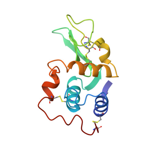
    Human lysozyme was co-crystallized with hexa-N-acetyl-chitohexaose, (GlcNAc)6, at pH 4.0 and 4.0 degrees C in a new orthorhombic form, where two protein molecules, MOL1 and MOL2, were contained in an asymmetric unit. The three-dimensional structure was refined to an R-factor of 17.0% at 1.6 A resolution. It was found that (GlcNAc)6 had already been cleaved to (GlcNAc)4 and (GlcNAc)2. In MOL1, (GlcNAc)4 was bound to the A, B, C, and D subsites, and binding sites of (GlcNAc)2 were close to the E and F subsites proposed on the basis of model building by Phillips and his colleagues. In MOL2, only the (GlcNAc)4 moiety could be found in the A, B, C and D subsites. Significant shifts of the backbone atoms were observed in the region of residues 102 to 120, which composed one side of the wall of the active site cleft. Consequently, the active cleft, with respect to the saccharide binding sites A, B and C, is narrower in both protein molecules. The residues 109 to 111 in site D of MOL1 are moved toward saccharide residue D, whereas those of MOL2 are only slightly shifted. In spite of these facts, the saccharide residues in site MOL1 and MOL2 are moved inside of the cleft. The distribution of water molecules and the hydrogen bond network in site D differ between the structures of MOL1 and MOL2. These structural changes in the active site cleft may be responsible for accommodating the substrate and releasing the products of hydrolysis. These results suggest that the three-dimensional structures of MOL1 and MOL2 remain in intermediate states between a transition state and an enzyme/product complex state.


  • Organizational Affiliation

    Protein Engineering Research Institute, Osaka, Japan.


Macromolecules
Find similar proteins by:  (by identity cutoff)  |  3D Structure
Entity ID: 1
MoleculeChains Sequence LengthOrganismDetailsImage
HUMAN LYSOZYME
A, B
130Homo sapiensMutation(s): 0 
EC: 3.2.1.17
UniProt & NIH Common Fund Data Resources
Find proteins for P61626 (Homo sapiens)
Explore P61626 
Go to UniProtKB:  P61626
PHAROS:  P61626
GTEx:  ENSG00000090382 
Entity Groups  
Sequence Clusters30% Identity50% Identity70% Identity90% Identity95% Identity100% Identity
UniProt GroupP61626
Sequence Annotations
Expand
  • Reference Sequence
Oligosaccharides

Help

Entity ID: 2
MoleculeChains Length2D Diagram Glycosylation3D Interactions
2-acetamido-2-deoxy-beta-D-glucopyranose-(1-4)-2-acetamido-2-deoxy-beta-D-glucopyranose-(1-4)-2-acetamido-2-deoxy-beta-D-glucopyranose-(1-4)-2-acetamido-2-deoxy-beta-D-glucopyranose
C, E
4N/A
Glycosylation Resources
GlyTouCan:  G01361SX
GlyCosmos:  G01361SX
GlyGen:  G01361SX
Entity ID: 3
MoleculeChains Length2D Diagram Glycosylation3D Interactions
2-acetamido-2-deoxy-beta-D-glucopyranose-(1-4)-2-acetamido-2-deoxy-beta-D-glucopyranose
D
2N/A
Glycosylation Resources
GlyTouCan:  G42666HT
GlyCosmos:  G42666HT
GlyGen:  G42666HT
Experimental Data & Validation

Experimental Data

  • Method: X-RAY DIFFRACTION
  • Resolution: 1.60 Å
  • R-Value Observed: 0.170 
  • Space Group: P 21 21 21
Unit Cell:
Length ( Å )Angle ( ˚ )
a = 46.99α = 90
b = 162.25β = 90
c = 32.52γ = 90
Software Package:
Software NamePurpose
PROLSQrefinement

Structure Validation

View Full Validation Report


Entry History 

Deposition Data

Revision History  (Full details and data files)

  • Version 1.0: 1995-04-20
    Type: Initial release
  • Version 1.1: 2008-03-24
    Changes: Version format compliance
  • Version 1.2: 2011-07-13
    Changes: Non-polymer description, Version format compliance
  • Version 1.3: 2017-11-29
    Changes: Derived calculations, Other
  • Version 2.0: 2020-07-29
    Type: Remediation
    Reason: Carbohydrate remediation
    Changes: Atomic model, Data collection, Derived calculations, Structure summary