Dissection of protein-carbohydrate interactions in mutant hen egg-white lysozyme complexes and their hydrolytic activity.
Maenaka, K., Matsushima, M., Song, H., Sunada, F., Watanabe, K., Kumagai, I.(1995) J Mol Biol 247: 281-293
- PubMed: 7707375
- DOI: https://doi.org/10.1006/jmbi.1994.0139
- Primary Citation of Related Structures:
1LZA, 1LZB, 1LZC, 1LZD, 1LZE, 1LZG - PubMed Abstract:
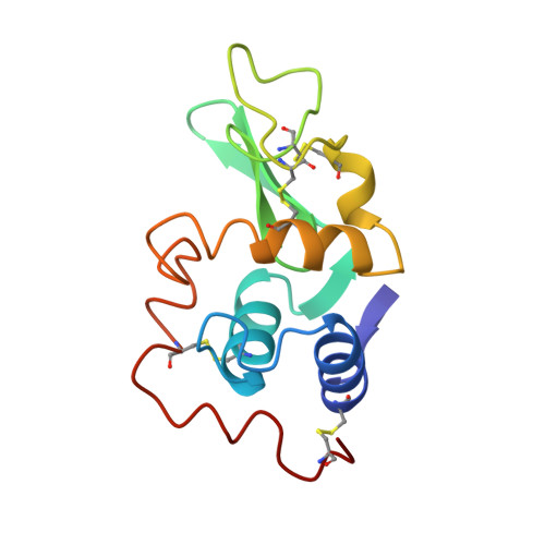
Trp62 in the binding subsite B of hen egg-white lysozyme shows general features often observed in protein-carbohydrate interactions including a stacking interaction and a hydrogen bonding network with water molecules. A previous report by our group showed that the perturbation of these interactions by substitution of Trp62 with tyrosine or phenylalanine affects the substrate binding modes and also enhances the hydrolytic activity. In order to elucidate the relationship between structural and functional changes of these protein-carbohydrate interactions, the Trp62Tyr and Trp62Phe mutants complexed with the substrate analogue, (GlcNAc)3, were analyzed at 1.8 A resolution by X-ray crystallography. The overall structures of the mutant enzymes are indistinguishable from that of the wild type enzyme. Although the wild-type enzyme binds (GlcNAc)3 in only one binding mode (A-B-C), the Trp62Tyr mutant binds (GlcNAc)3 in two binding modes (A-B-C, B-C-D) and the Trp62Phe mutant has an even weaker binding mode. The aromatic rings of Tyr62 and Phe62 maintain their interactions with the carbohydrate molecules, but make fewer stacking interactions with the GlcNAc in the B site than the wild-type enzyme does. The hydroxyl group of Tyr62 interacts weakly with a water molecule which mediates hydrogen bonding in the GlcNAc residues in the B and C sites. The C-6 hydroxyl group of the GlcNAc residue in the C site rotates around the C-5-C-6 bond to complete the hydrogen bond network in the Trp62Tyr mutant-(GlcNAc)3 complex. On the other hand, this hydrogen bonding network does not form in the Trp62Phe mutant-(GlcNAc)3. In addition to these structural studies, the kinetic parameters of the hydrolysis of 4-methylumbelliferyl N-acetyl-chitotriose, ((GlcNAc)3-MeU), have been determined in order to further characterize the enzymatic properties of these mutant lysozymes. This demonstrates that the modulation of the hydrogen bonding network, including the flexible part of the carbohydrate and water molecules and/or the slight reduction of stacking interaction in the B site, alters the binding mode toward the carbohydrate and induces an enhancement of the hydrolytic activity.
Organizational Affiliation:
Department of Chemistry and Biotechnology, Faculty of Engineering, The University of Tokyo, Japan.