Structural and functional analyses of the Arg-Gly-Asp sequence introduced into human lysozyme.
Yamada, T., Matsushima, M., Inaka, K., Ohkubo, T., Uyeda, A., Maeda, T., Titani, K., Sekiguchi, K., Kikuchi, M.(1993) J Biol Chem 268: 10588-10592
- PubMed: 8486712
- Primary Citation of Related Structures:
1LZ5, 1LZ6 - PubMed Abstract:
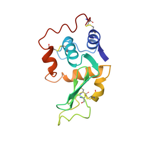
To determine the functional conformation of the Arg-Gly-Asp (RGD) sequence, we have constructed mutant proteins by inserting 4-12 amino acid residues from the RGD region of human fibronectin between Val74 and Asn75 of human lysozyme. RGDS-, GRGDSP-, TGRGDSPA-, VTGRGDSPAS-, and AVTGRGDS-PASS-introduced mutant lysozymes were expressed in yeast, purified, and designated as RGD4, -6, -8, -10, and -12, respectively. Using baby hamster kidney cells, RGD8, RGD10, and RGD12 were shown to possess high cell adhesion activity nearly equal to 10% of human vitronectin activity. RGD4 and RGD6 exhibited somewhat lower cell adhesion activity. The activities of these mutant proteins were inhibited by the addition of either GRGDSP peptide or polyclonal antibody against vitronectin receptor, as was the case for the vitronectin activity. The results suggest that the cell adhesion signals are transduced to cells through the interaction with the vitronectin receptor. The three-dimensional structures of RGD4 and RGD8 were determined at 1.8-A resolution by x-ray crystallography. A model of the inserted region in RGD4 could be built in the electron density map, but the positions of the preceding residues, Ala73-Val74, were uncertain. The inserted region in RGD8 did not demonstrate continuous electron densities. The results suggest that these RGD sequence-containing regions are highly flexible and that such flexibility could allow the conformation of the RGD regions to be induced to fit into the binding pocket of the integrin receptor.
Organizational Affiliation:
Protein Engineering Research Institute, Osaka, Japan.