1LP1

Protein Z in complex with an in vitro selected affibody


Experimental Data Snapshot

  • Method: X-RAY DIFFRACTION
  • Resolution: 2.30 Å
  • R-Value Free: 0.256 
  • R-Value Work: 0.224 
  • R-Value Observed: 0.225 

wwPDB Validation   3D Report Full Report


This is version 1.5 of the entry. See complete history


Literature

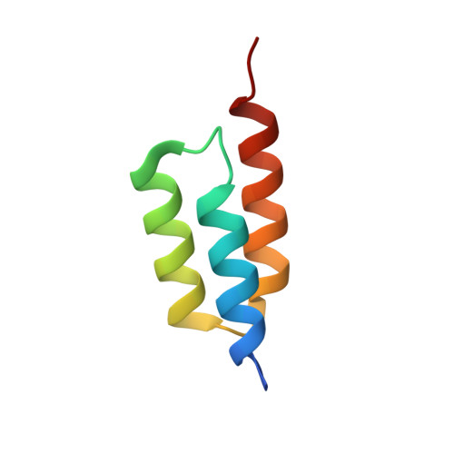
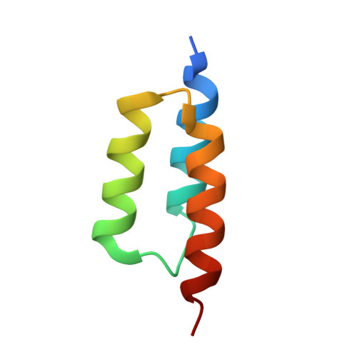
Structural basis for recognition by an in vitro evolved affibody.

Hogbom, M.Eklund, M.Nygren, P.A.Nordlund, P.

(2003) Proc Natl Acad Sci U S A 100: 3191-3196

  • DOI: https://doi.org/10.1073/pnas.0436100100
  • Primary Citation of Related Structures:  
    1LP1

  • PubMed Abstract: 

    The broad binding repertoire of antibodies has permitted their use in a wide range of applications. However, some uses of antibodies are precluded due to limitations in the efficiency of antibody generation. In vitro evolved binding proteins, selected from combinatorial libraries generated around various alternative structural scaffolds, are promising alternatives to antibodies. We have solved the crystal structure of a complex of an all alpha-helical in vitro selected binding protein (affibody) bound to protein Z, an IgG Fc-binding domain derived from staphylococcal protein A. The structure of the complex reveals an extended and complementary binding surface with similar properties to protein-antibody interactions. The surface region of protein Z recognized by the affibody is strikingly similar to the one used for IgG(1) Fc binding, suggesting that this surface contains potential hot-spots for binding. The implications of the selected affibody binding-mode for its application as a universal binding protein are discussed.


  • Organizational Affiliation

    Department of Biochemistry and Biophysics, Stockholm University, Roslagstullsbacken 15, Albanova University Center, SE-10691 Stockholm, Sweden.


Macromolecules
Find similar proteins by:  (by identity cutoff)  |  3D Structure
Entity ID: 1
MoleculeChains Sequence LengthOrganismDetailsImage
Affibody binding protein Z58Staphylococcus aureusMutation(s): 0 
Entity Groups  
Sequence Clusters30% Identity50% Identity70% Identity90% Identity95% Identity100% Identity
Sequence Annotations
Expand
  • Reference Sequence
Find similar proteins by:  (by identity cutoff)  |  3D Structure
Entity ID: 2
MoleculeChains Sequence LengthOrganismDetailsImage
Immunoglobulin G binding protein A58Staphylococcus aureusMutation(s): 2 
UniProt
Find proteins for P38507 (Staphylococcus aureus)
Explore P38507 
Go to UniProtKB:  P38507
Entity Groups  
Sequence Clusters30% Identity50% Identity70% Identity90% Identity95% Identity100% Identity
UniProt GroupP38507
Sequence Annotations
Expand
  • Reference Sequence
Experimental Data & Validation

Experimental Data

  • Method: X-RAY DIFFRACTION
  • Resolution: 2.30 Å
  • R-Value Free: 0.256 
  • R-Value Work: 0.224 
  • R-Value Observed: 0.225 
  • Space Group: P 61 2 2
Unit Cell:
Length ( Å )Angle ( ˚ )
a = 55.546α = 90
b = 55.546β = 90
c = 155.749γ = 120
Software Package:
Software NamePurpose
DENZOdata reduction
SCALEPACKdata scaling
EPMRphasing
CNSrefinement

Structure Validation

View Full Validation Report


Entry History 

Deposition Data

Revision History  (Full details and data files)

  • Version 1.0: 2003-03-18
    Type: Initial release
  • Version 1.1: 2008-04-28
    Changes: Version format compliance
  • Version 1.2: 2011-07-13
    Changes: Version format compliance
  • Version 1.3: 2018-05-23
    Changes: Data collection
  • Version 1.4: 2021-11-10
    Changes: Database references, Derived calculations
  • Version 1.5: 2023-10-25
    Changes: Data collection, Refinement description