Protein hydration and water structure: X-ray analysis of a closely packed protein crystal with very low solvent content.
Madhusudan, Kodandapani, R., Vijayan, M.(1993) Acta Crystallogr D Biol Crystallogr 49: 234-245
- PubMed: 15299529
- DOI: https://doi.org/10.1107/S090744499200653X
- Primary Citation of Related Structures:
1LMA - PubMed Abstract:
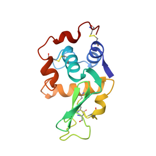
Low-humidity monoclinic lysozyme, resulting from a water-mediated transformation, has one of the lowest solvent contents (22% by volume) observed in a protein crystal. Its structure has been solved by the molecular replacement method and refined to an R value of 0.175 for 7684 observed reflections in the 10-1.75 A resolution shell. 90% of the solvent in the well ordered crystals could be located. Favourable sites of hydration on the protein surface include side chains with multiple hydrogen-bonding centres, and regions between short hydrophilic side chains and the main-chain CO or NH groups of the same or nearby residues. Major secondary structural features are not disrupted by hydration. However, the free CO groups at the C terminii and, to a lesser extent, the NH groups at the N terminii of helices provide favourable sites for water interactions, as do reverse turns and regions which connect beta-structure and helices. The hydration shell consists of discontinuous networks of water molecules, the maximum number of molecules in a network being ten. The substrate-binding cleft is heavily hydrated, as is the main loop region which is stabilized by water interactions. The protein molecules are close packed in the crystals with a molecular coordination number of 14. Arginyl residues are extensively involved in intermolecular hydrogen bonds and water bridges. The water molecules in the crystal are organized into discrete clusters. A distinctive feature of the clusters is the frequent occurrence of three-membered rings. The protein molecules undergo substantial rearrangement during the transformation from the native to the low-humidity form. The main-chain conformations in the two forms are nearly the same, but differences exist in the side-chain conformation. The differences are particularly pronounced in relation to Trp 62 and Trp 63. The shift in Trp 62 is especially interesting as it is also known to move during inhibitor binding.
Organizational Affiliation:
Molecular Biophysics Unit, Indian Institute of Science, Bangalore, India.