Comparison of the three-dimensional structures of a human Bence-Jones dimer crystallized on Earth and aboard US Space Shuttle Mission STS-95
Terzyan, S.S., DeWitt, C.R., Ramsland, P.A., Bourne, P.C., Edmundson, A.B.(2003) J Mol Recognit 16: 83-90
- PubMed: 12720277
- DOI: https://doi.org/10.1002/jmr.610
- Primary Citation of Related Structures:
1LGV, 1LHZ - PubMed Abstract:
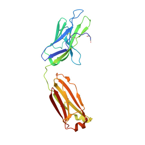
Crystals of a human (Sea) Bence-Jones dimer were produced in a capillary by vapor diffusion under microgravity conditions in the 9 day US Space Shuttle Mission STS-95. In comparison to ground-based experiments, nucleation was facile and spontaneous in space. Appearance of a very large (8 x 1.6 x 1.0 mm) crystal in a short time period is a strong endorsement for the use of microgravity to produce crystals sufficiently large for neutron diffraction studies. The Sea dimer crystallized in the orthorhombic space group P2(1)2(1)2(1), with a = 48.9 A, b = 85.2 A, and c = 114.0 A. The crystals grown in microgravity exhibited significantly lower mosaicities than those of ground-based crystals and the X-ray diffraction data had a lower overall B factor. Three-dimensional structures determined by X-ray analysis at two temperatures (100 and 293 K) were indistinguishable from those obtained from ground-based crystals. However, both the crystallographic R factor and the free R factor were slightly lower in the models derived from crystals produced in microgravity. The major difference between the two crystal growth systems is a lack of convection and sedimentation in a microgravity environment. This environment resulted in the growth of much larger, higher-quality crystals of the Sea Bence-Jones protein. Structurally, heretofore unrecognized grooves on the external surfaces of the Sea and other immunoglobulin-derived fragments are regular features and may offer supplementary binding regions for super antigens and other elongated ligands in the bloodstream and perivascular tissues.
Organizational Affiliation:
Crystallography Program, Oklahoma Medical Research Foundation, 825 N.E. 13th Street, Oklahoma City, OK 73104, USA.