1LFA

CD11A I-DOMAIN WITH BOUND MN++


Experimental Data Snapshot

  • Method: X-RAY DIFFRACTION
  • Resolution: 1.80 Å
  • R-Value Free: 0.251 
  • R-Value Work: 0.190 
  • R-Value Observed: 0.190 

wwPDB Validation   3D Report Full Report


This is version 1.3 of the entry. See complete history


Literature

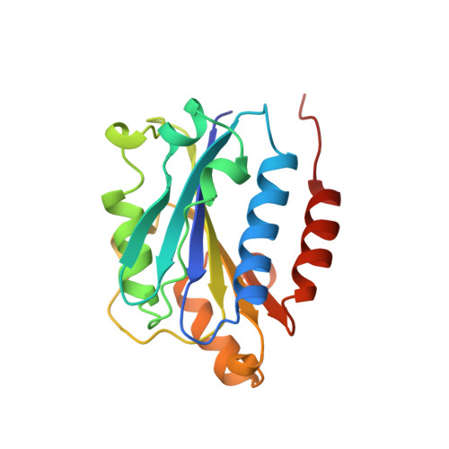
Crystal structure of the I-domain from the CD11a/CD18 (LFA-1, alpha L beta 2) integrin.

Qu, A.Leahy, D.J.

(1995) Proc Natl Acad Sci U S A 92: 10277-10281

  • DOI: https://doi.org/10.1073/pnas.92.22.10277
  • Primary Citation of Related Structures:  
    1LFA

  • PubMed Abstract: 

    We report the 1.8-A crystal structure of the CD11a I-domain with bound manganese ion. The CD11a I-domain contains binding sites for intercellular adhesion molecules 1 and 3 and can exist in both low- and high-affinity states. The metal-bound form reported here is likely to represent a high-affinity state. The CD11a I-domain structure reveals a strained hydrophobic ridge adjacent to the bound metal ion that may serve as a ligand-binding surface and is likely to rearrange in the absence of bound metal ion. The CD11a I-domain is homologous to domains found in von Willebrand factor, and mapping of mutations found in types 2a and 2b von Willebrand disease onto this structure allows consideration of the molecular basis of these forms of the disease.


  • Organizational Affiliation

    Department of Biophysics and Biophysical Chemistry, Johns Hopkins University School of Medicine, Baltimore, MD 21205, USA.


Macromolecules
Find similar proteins by:  (by identity cutoff)  |  3D Structure
Entity ID: 1
MoleculeChains Sequence LengthOrganismDetailsImage
CD11A
A, B
187Homo sapiensMutation(s): 0 
UniProt & NIH Common Fund Data Resources
Find proteins for P20701 (Homo sapiens)
Explore P20701 
Go to UniProtKB:  P20701
PHAROS:  P20701
GTEx:  ENSG00000005844 
Entity Groups  
Sequence Clusters30% Identity50% Identity70% Identity90% Identity95% Identity100% Identity
UniProt GroupP20701
Sequence Annotations
Expand
  • Reference Sequence
Experimental Data & Validation

Experimental Data

  • Method: X-RAY DIFFRACTION
  • Resolution: 1.80 Å
  • R-Value Free: 0.251 
  • R-Value Work: 0.190 
  • R-Value Observed: 0.190 
  • Space Group: C 1 2 1
Unit Cell:
Length ( Å )Angle ( ˚ )
a = 131.13α = 90
b = 45.45β = 99.8
c = 66.13γ = 90
Software Package:
Software NamePurpose
X-PLORmodel building
X-PLORrefinement
X-PLORphasing

Structure Validation

View Full Validation Report


Entry History 

Deposition Data

Revision History  (Full details and data files)

  • Version 1.0: 1996-01-29
    Type: Initial release
  • Version 1.1: 2008-03-24
    Changes: Version format compliance
  • Version 1.2: 2011-07-13
    Changes: Version format compliance
  • Version 1.3: 2024-02-14
    Changes: Data collection, Database references, Derived calculations, Other