1KW4

Polyhomeotic SAM domain structure


Experimental Data Snapshot

  • Method: X-RAY DIFFRACTION
  • Resolution: 1.75 Å
  • R-Value Free: 0.234 
  • R-Value Work: 0.220 
  • R-Value Observed: 0.220 

wwPDB Validation   3D Report Full Report


This is version 1.3 of the entry. See complete history


Literature

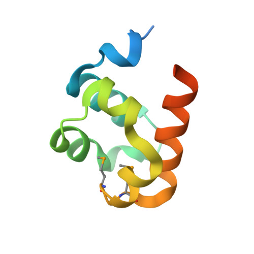
The SAM domain of polyhomeotic forms a helical polymer.

Kim, C.A.Gingery, M.Pilpa, R.M.Bowie, J.U.

(2002) Nat Struct Biol 9: 453-457

  • DOI: https://doi.org/10.1038/nsb802
  • Primary Citation of Related Structures:  
    1KW4

  • PubMed Abstract: 

    The polycomb group (PcG) proteins are important in the maintenance of stable repression patterns during development. Several PcG members contain a protein protein interaction module called a SAM domain (also known as SPM, PNT and HLH). Here we report the high-resolution structure of the SAM domain of polyhomeotic (Ph). Ph-SAM forms a helical polymer structure, providing a likely mechanism for the extension of PcG complexes. The structure of the polymer resembles that formed by the SAM domain of another transcriptional repressor, TEL. The formation of these polymer structures by SAM domains in two divergent repressors suggests a conserved mode of repression involving a higher order chromatin structure.


  • Organizational Affiliation

    Department of Chemistry and Biochemistry, Laboratory of Structural Biology and Molecular Medicine, Molecular Biology Institute, UCLA, 611 Charles E. Young Drive East, Los Angeles, California 90095-1570, USA.


Macromolecules
Find similar proteins by:  (by identity cutoff)  |  3D Structure
Entity ID: 1
MoleculeChains Sequence LengthOrganismDetailsImage
Polyhomeotic89Drosophila melanogasterMutation(s): 3 
UniProt
Find proteins for P39769 (Drosophila melanogaster)
Explore P39769 
Go to UniProtKB:  P39769
Entity Groups  
Sequence Clusters30% Identity50% Identity70% Identity90% Identity95% Identity100% Identity
UniProt GroupP39769
Sequence Annotations
Expand
  • Reference Sequence
Small Molecules
Modified Residues  1 Unique
IDChains TypeFormula2D DiagramParent
MSE
Query on MSE
A
L-PEPTIDE LINKINGC5 H11 N O2 SeMET
Experimental Data & Validation

Experimental Data

  • Method: X-RAY DIFFRACTION
  • Resolution: 1.75 Å
  • R-Value Free: 0.234 
  • R-Value Work: 0.220 
  • R-Value Observed: 0.220 
  • Space Group: P 65
Unit Cell:
Length ( Å )Angle ( ˚ )
a = 59.836α = 90
b = 59.836β = 90
c = 44.896γ = 120
Software Package:
Software NamePurpose
SOLVEphasing
CNSrefinement
DENZOdata reduction
SCALEPACKdata scaling

Structure Validation

View Full Validation Report


Entry History 

Deposition Data

Revision History  (Full details and data files)

  • Version 1.0: 2002-06-05
    Type: Initial release
  • Version 1.1: 2008-04-27
    Changes: Version format compliance
  • Version 1.2: 2011-07-13
    Changes: Version format compliance
  • Version 1.3: 2021-10-27
    Changes: Database references, Derived calculations