1KTR

Crystal Structure of the Anti-His Tag Antibody 3D5 Single-Chain Fragment (scFv) in Complex with a Oligohistidine peptide


Experimental Data Snapshot

  • Method: X-RAY DIFFRACTION
  • Resolution: 2.70 Å
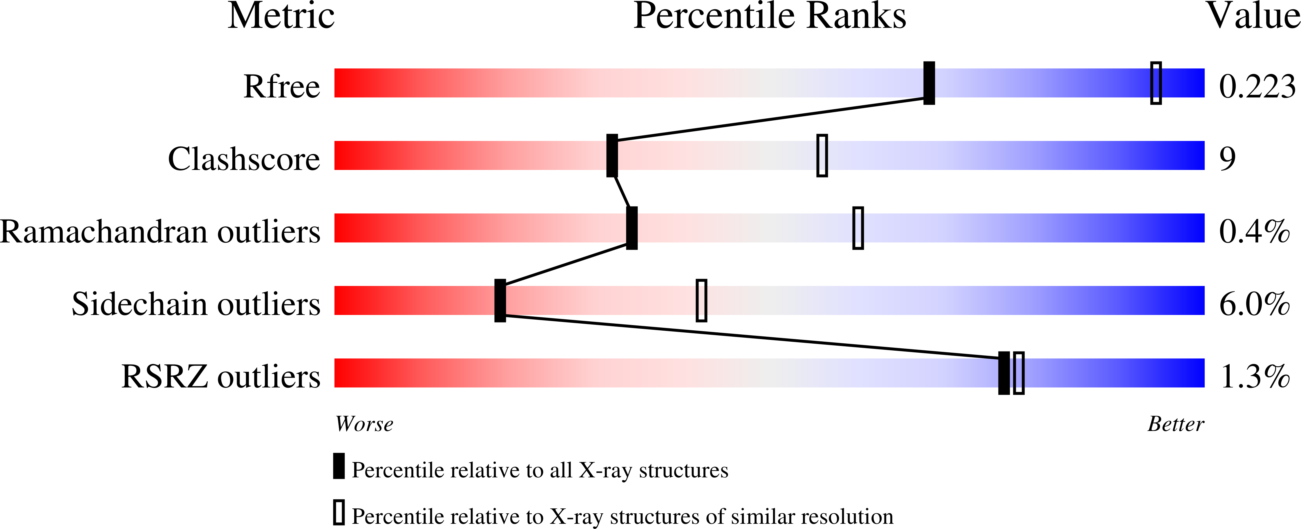
  • R-Value Free: 0.222 
  • R-Value Work: 0.190 

wwPDB Validation   3D Report Full Report


This is version 2.1 of the entry. See complete history


Literature

Crystal structure of the anti-His tag antibody 3D5 single-chain fragment complexed to its antigen.

Kaufmann, M.Lindner, P.Honegger, A.Blank, K.Tschopp, M.Capitani, G.Pluckthun, A.Grutter, M.G.

(2002) J Mol Biol 318: 135-147

  • DOI: https://doi.org/10.1016/S0022-2836(02)00038-4
  • Primary Citation of Related Structures:  
    1KTR

  • PubMed Abstract: 

    The crystal structure of a mutant form of the single-chain fragment (scFv), derived from the monoclonal anti-His tag antibody 3D5, in complex with a hexahistidine peptide has been determined at 2.7 A resolution. The peptide binds to a deep pocket formed at the interface of the variable domains of the light and the heavy chain, mainly through hydrophobic interaction to aromatic residues and hydrogen bonds to acidic residues. The antibody recognizes the C-terminal carboxylate group of the peptide as well as the main chain of the last four residues and the last three imidazole side-chains. The crystals have a solvent content of 77% (v/v) and form 70 A-wide channels that would allow the diffusion of peptides or even small proteins. The anti-His scFv crystals could thus act as a framework for the crystallization of His-tagged target proteins. Designed mutations in framework regions of the scFv lead to high-level expression of soluble protein in the periplasm of Escherichia coli. The recombinant anti-His scFv is a convenient detection tool when fused to alkaline phosphatase. When immobilized on a matrix, the antibody can be used for affinity purification of recombinant proteins carrying a very short tag of just three histidine residues, suitable for crystallization. The experimental structure is now the basis for the design of antibodies with even higher stability and affinity.


  • Organizational Affiliation

    Biochemisches Institut, Universität Zürich, Winterthurerstrasse 190, CH-8057 Zurich, Switzerland.


Macromolecules
Find similar proteins by:  (by identity cutoff)  |  3D Structure
Entity ID: 1
MoleculeChains Sequence LengthOrganismDetailsImage
Anti-his tag antibody 3d5 variable light chain, Peptide linker, Anti-his tag antibody 3d5 variable heavy chainA [auth L]250Mus musculusMutation(s): 0 
Gene Names: Monoclonal Antibody 3D5
Entity Groups  
Sequence Clusters30% Identity50% Identity70% Identity90% Identity95% Identity100% Identity
Sequence Annotations
Expand
  • Reference Sequence

Find similar proteins by:  Sequence   |   3D Structure  

Entity ID: 2
MoleculeChains Sequence LengthOrganismDetailsImage
Oligohistidine peptide AntigenB [auth P]6synthetic constructMutation(s): 0 
Sequence Annotations
Expand
  • Reference Sequence
Experimental Data & Validation

Experimental Data

  • Method: X-RAY DIFFRACTION
  • Resolution: 2.70 Å
  • R-Value Free: 0.222 
  • R-Value Work: 0.190 
  • Space Group: P 32 2 1
Unit Cell:
Length ( Å )Angle ( ˚ )
a = 106.51α = 90
b = 106.51β = 90
c = 92.8γ = 120
Software Package:
Software NamePurpose
DENZOdata reduction
SCALEPACKdata scaling
AMoREphasing
CNSrefinement

Structure Validation

View Full Validation Report


Entry History 

Deposition Data

Revision History  (Full details and data files)

  • Version 1.0: 2002-05-15
    Type: Initial release
  • Version 1.1: 2008-04-27
    Changes: Version format compliance
  • Version 1.2: 2011-07-13
    Changes: Version format compliance
  • Version 2.0: 2018-05-02
    Changes: Advisory, Atomic model, Data collection, Database references, Derived calculations, Polymer sequence, Source and taxonomy, Structure summary
  • Version 2.1: 2023-08-16
    Changes: Data collection, Database references, Derived calculations, Refinement description