1KR7

Crystal structure of the nerve tissue mini-hemoglobin from the nemertean worm Cerebratulus lacteus


Experimental Data Snapshot

  • Method: X-RAY DIFFRACTION
  • Resolution: 1.50 Å
  • R-Value Free: 0.187 
  • R-Value Work: 0.153 
  • R-Value Observed: 0.156 

wwPDB Validation   3D Report Full Report


Ligand Structure Quality Assessment 


This is version 1.3 of the entry. See complete history


Literature

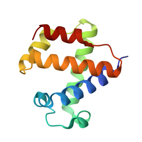
The 109 residue nerve tissue minihemoglobin from Cerebratulus lacteus highlights striking structural plasticity of the alpha-helical globin fold

Pesce, A.Nardini, M.Dewilde, S.Geuens, E.Yamauchi, k.Ascenzi, P.Riggs, A.F.Moens, L.Bolognesi, M.

(2002) Structure 10: 725-735

  • DOI: https://doi.org/10.1016/s0969-2126(02)00763-3
  • Primary Citation of Related Structures:  
    1KR7

  • PubMed Abstract: 

    A very short hemoglobin (CerHb; 109 amino acids) binds O(2) cooperatively in the nerve tissue of the nemertean worm Cerebratulus lacteus to sustain neural activity during anoxia. Sequence analysis suggests that CerHb tertiary structure may be unique among the known globin fold evolutionary variants. The X-ray structure of oxygenated CerHb (R factor 15.3%, at 1.5 A resolution) displays deletion of the globin N-terminal A helix, an extended GH region, a very short H helix, and heme solvent shielding based on specific aromatic residues. The heme-bound O(2) is stabilized by hydrogen bonds to the distal TyrB10-GlnE7 pair. Ligand access to heme may take place through a wide protein matrix tunnel connecting the distal site to a surface cleft located between the E and H helices.


  • Organizational Affiliation

    Department of Physics, INFM, Advanced Biotechnology Centre, University of Genova, Largo Rosanna Benzi 10, I-16146 Genova, Italy.


Macromolecules
Find similar proteins by:  (by identity cutoff)  |  3D Structure
Entity ID: 1
MoleculeChains Sequence LengthOrganismDetailsImage
Neural globin110Cerebratulus lacteusMutation(s): 0 
UniProt
Find proteins for O76242 (Cerebratulus lacteus)
Explore O76242 
Go to UniProtKB:  O76242
Entity Groups  
Sequence Clusters30% Identity50% Identity70% Identity90% Identity95% Identity100% Identity
UniProt GroupO76242
Sequence Annotations
Expand
  • Reference Sequence
Experimental Data & Validation

Experimental Data

  • Method: X-RAY DIFFRACTION
  • Resolution: 1.50 Å
  • R-Value Free: 0.187 
  • R-Value Work: 0.153 
  • R-Value Observed: 0.156 
  • Space Group: P 21 21 21
Unit Cell:
Length ( Å )Angle ( ˚ )
a = 42.721α = 90
b = 43.173β = 90
c = 60.11γ = 90
Software Package:
Software NamePurpose
SOLVEphasing
REFMACrefinement
DENZOdata reduction
SCALEPACKdata scaling

Structure Validation

View Full Validation Report


Ligand Structure Quality Assessment 


Entry History 

Deposition Data

Revision History  (Full details and data files)

  • Version 1.0: 2002-05-15
    Type: Initial release
  • Version 1.1: 2007-10-16
    Changes: Version format compliance
  • Version 1.2: 2011-07-13
    Changes: Version format compliance
  • Version 1.3: 2024-02-14
    Changes: Data collection, Database references, Derived calculations