1KIG

BOVINE FACTOR XA


Experimental Data Snapshot

  • Method: X-RAY DIFFRACTION
  • Resolution: 3.00 Å
  • R-Value Work: 0.187 
  • R-Value Observed: 0.187 

wwPDB Validation   3D Report Full Report


This is version 1.2 of the entry. See complete history


Literature

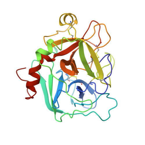
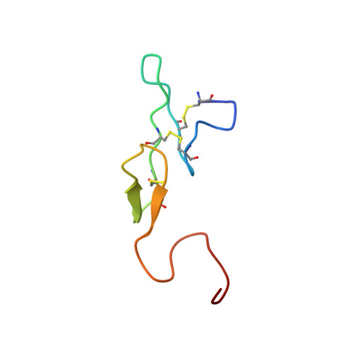
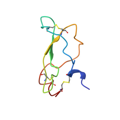
Unexpected binding mode of tick anticoagulant peptide complexed to bovine factor Xa.

Wei, A.Alexander, R.S.Duke, J.Ross, H.Rosenfeld, S.A.Chang, C.H.

(1998) J Mol Biol 283: 147-154

  • DOI: https://doi.org/10.1006/jmbi.1998.2069
  • Primary Citation of Related Structures:  
    1KIG

  • PubMed Abstract: 

    The structure of recombinant tick anticoagulant peptide (rTAP) complexed to bovine factor Xa at 3.0 A resolution reveals the structural basis for the specificity and the high affinity of rTAP. Three N-terminal residues, Tyr501, Asn502 and Arg503, play a critical role in the complex formation as suggested by earlier mutagenic studies and the ornithodorin-thrombin complex. Unexpectedly, the side-chain of Tyr501 is located in the S1 pocket, although factor Xa favors arginine as a P1 residue. Arg503 is located at the aryl binding pocket and forms a salt-bridge with Glu97 of factor Xa. The autolysis loop, which is disordered in the uninhibited factor Xa structure, is involved in the formation of the complex as a part of the secondary binding site. The C-terminal helix of rTAP interacts with factor Xa as a secondary binding determinant. The N-terminal residues of rTAP reorganize during the formation of the factor Xa-rTAP complex from the conformation found in the solution into an extended conformation. The presence of the secondary binding site confirms the proposed two-step kinetic mechanism based on the results of a mutagenesis study.


  • Organizational Affiliation

    Department of Chemical and Physical Sciences, The DuPont Pharmaceuticals Company, Wilmington DE 19880, USA.


Macromolecules
Find similar proteins by:  (by identity cutoff)  |  3D Structure
Entity ID: 1
MoleculeChains Sequence LengthOrganismDetailsImage
FACTOR XAA [auth H]241Bos taurusMutation(s): 0 
EC: 3.4.21.6
UniProt
Find proteins for P00743 (Bos taurus)
Explore P00743 
Go to UniProtKB:  P00743
Entity Groups  
Sequence Clusters30% Identity50% Identity70% Identity90% Identity95% Identity100% Identity
UniProt GroupP00743
Sequence Annotations
Expand
  • Reference Sequence
Find similar proteins by:  (by identity cutoff)  |  3D Structure
Entity ID: 2
MoleculeChains Sequence LengthOrganismDetailsImage
FACTOR XAB [auth L]51Bos taurusMutation(s): 0 
EC: 3.4.21.6
UniProt
Find proteins for P00743 (Bos taurus)
Explore P00743 
Go to UniProtKB:  P00743
Entity Groups  
Sequence Clusters30% Identity50% Identity70% Identity90% Identity95% Identity100% Identity
UniProt GroupP00743
Sequence Annotations
Expand
  • Reference Sequence
Find similar proteins by:  (by identity cutoff)  |  3D Structure
Entity ID: 3
MoleculeChains Sequence LengthOrganismDetailsImage
ANTICOAGULANT PEPTIDEC [auth I]60Ornithodoros moubataMutation(s): 0 
UniProt
Find proteins for P17726 (Ornithodoros moubata)
Explore P17726 
Go to UniProtKB:  P17726
Entity Groups  
Sequence Clusters30% Identity50% Identity70% Identity90% Identity95% Identity100% Identity
UniProt GroupP17726
Sequence Annotations
Expand
  • Reference Sequence
Experimental Data & Validation

Experimental Data

  • Method: X-RAY DIFFRACTION
  • Resolution: 3.00 Å
  • R-Value Work: 0.187 
  • R-Value Observed: 0.187 
  • Space Group: P 42 21 2
Unit Cell:
Length ( Å )Angle ( ˚ )
a = 133.1α = 90
b = 133.1β = 90
c = 68.8γ = 90
Software Package:
Software NamePurpose
X-PLORmodel building
X-PLORrefinement
X-PLORphasing

Structure Validation

View Full Validation Report


Entry History 

Deposition Data

Revision History  (Full details and data files)

  • Version 1.0: 1998-10-28
    Type: Initial release
  • Version 1.1: 2008-03-24
    Changes: Version format compliance
  • Version 1.2: 2011-07-13
    Changes: Version format compliance