1K3S

Type III Secretion Chaperone SigE


Experimental Data Snapshot

  • Method: X-RAY DIFFRACTION
  • Resolution: 1.90 Å
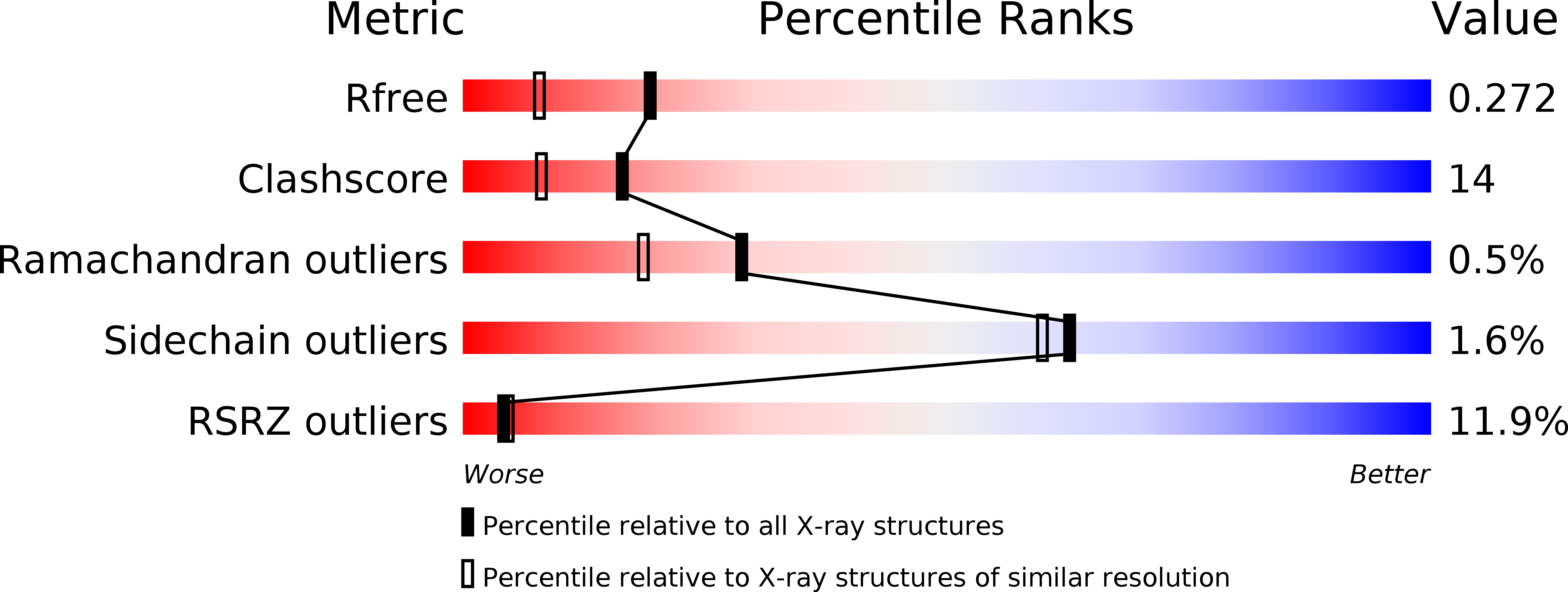
  • R-Value Free: 0.272 
  • R-Value Work: 0.224 
  • R-Value Observed: 0.224 

wwPDB Validation   3D Report Full Report


This is version 1.3 of the entry. See complete history


Literature

Structural and biochemical characterization of the type III secretion chaperones CesT and SigE.

Luo, Y.Bertero, M.G.Frey, E.A.Pfuetzner, R.A.Wenk, M.R.Creagh, L.Marcus, S.L.Lim, D.Sicheri, F.Kay, C.Haynes, C.Finlay, B.B.Strynadka, N.C.

(2001) Nat Struct Biol 8: 1031-1036

  • DOI: https://doi.org/10.1038/nsb717
  • Primary Citation of Related Structures:  
    1K3E, 1K3S

  • PubMed Abstract: 

    Several Gram-negative bacterial pathogens have evolved a type III secretion system to deliver virulence effector proteins directly into eukaryotic cells, a process essential for disease. This specialized secretion process requires customized chaperones specific for particular effector proteins. The crystal structures of the enterohemorrhagic Escherichia coli O157:H7 Tir-specific chaperone CesT and the Salmonella enterica SigD-specific chaperone SigE reveal a common overall fold and formation of homodimers. Site-directed mutagenesis suggests that variable, delocalized hydrophobic surfaces observed on the chaperone homodimers are responsible for specific binding to a particular effector protein. Isothermal titration calorimetry studies of Tir-CesT and enzymatic activity profiles of SigD-SigE indicate that the effector proteins are not globally unfolded in the presence of their cognate chaperones.


  • Organizational Affiliation

    Department of Biochemistry and Molecular Biology, University of British Columbia, Vancouver V6T 1Z3, Canada.


Macromolecules
Find similar proteins by:  (by identity cutoff)  |  3D Structure
Entity ID: 1
MoleculeChains Sequence LengthOrganismDetailsImage
SigE
A, B
113Salmonella entericaMutation(s): 3 
UniProt
Find proteins for A0A0F6AZQ4 (Salmonella typhimurium (strain 14028s / SGSC 2262))
Explore A0A0F6AZQ4 
Go to UniProtKB:  A0A0F6AZQ4
Entity Groups  
Sequence Clusters30% Identity50% Identity70% Identity90% Identity95% Identity100% Identity
UniProt GroupA0A0F6AZQ4
Sequence Annotations
Expand
  • Reference Sequence
Small Molecules
Ligands 1 Unique
IDChains Name / Formula / InChI Key2D Diagram3D Interactions
PO4
Query on PO4

Download Ideal Coordinates CCD File 
C [auth A]PHOSPHATE ION
O4 P
NBIIXXVUZAFLBC-UHFFFAOYSA-K
Modified Residues  1 Unique
IDChains TypeFormula2D DiagramParent
MSE
Query on MSE
A, B
L-PEPTIDE LINKINGC5 H11 N O2 SeMET
Experimental Data & Validation

Experimental Data

  • Method: X-RAY DIFFRACTION
  • Resolution: 1.90 Å
  • R-Value Free: 0.272 
  • R-Value Work: 0.224 
  • R-Value Observed: 0.224 
  • Space Group: P 21 21 21
Unit Cell:
Length ( Å )Angle ( ˚ )
a = 44.497α = 90
b = 48.725β = 90
c = 94.532γ = 90
Software Package:
Software NamePurpose
SOLVEphasing
CNSrefinement
DENZOdata reduction
SCALEPACKdata scaling

Structure Validation

View Full Validation Report


Entry History 

Deposition Data

Revision History  (Full details and data files)

  • Version 1.0: 2001-11-28
    Type: Initial release
  • Version 1.1: 2008-04-27
    Changes: Version format compliance
  • Version 1.2: 2011-07-13
    Changes: Version format compliance
  • Version 1.3: 2016-05-25
    Changes: Source and taxonomy