1JYI

CONCANAVALIN A/12-MER PEPTIDE COMPLEX


Experimental Data Snapshot

  • Method: X-RAY DIFFRACTION
  • Resolution: 2.75 Å
  • R-Value Free: 0.265 
  • R-Value Work: 0.192 

wwPDB Validation   3D Report Full Report


This is version 1.4 of the entry. See complete history


Literature

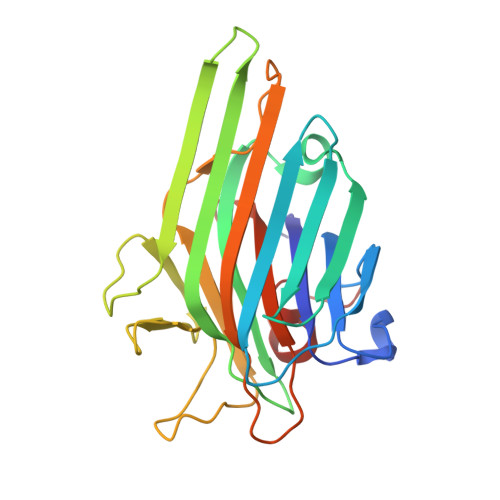
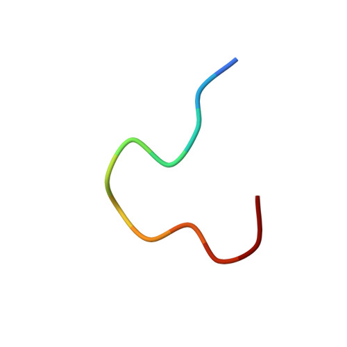
Structural and Functional Consequences of Peptide-carbohydrate Mimicry. Crystal Structure of a Carbohydrate-mimicking Peptide Bound to Concanavalin A.

Jain, D.Kaur, K.J.Sundaravadivel, B.Salunke, D.M.

(2000) J Biol Chem 275: 16098-16102

  • DOI: https://doi.org/10.1074/jbc.275.21.16098
  • Primary Citation of Related Structures:  
    1JYI

  • PubMed Abstract: 

    The functional consequences of peptide-carbohydrate mimicry were analyzed on the basis of the crystal structure of concanavalin A (ConA) in complex with a carbohydrate-mimicking peptide, DVFYPYPYASGS. The peptide binds to the non-crystallographically related monomers of two independent dimers of ConA in two different modes, in slightly different conformations, demonstrating structural adaptability in ConA-peptide recognition. In one mode, the peptide has maximum interactions with ConA, and in the other, it shows relatively fewer contacts within this site but significant contacts with the symmetry-related subunit. Neither of the peptide binding sites overlaps with the structurally characterized mannose and trimannose binding sites on ConA. Despite this, the functional mimicry between the peptide and carbohydrate ligands was evident. The peptide-inhibited ConA induced T cell proliferation in a dose-dependent manner. The effect of the designed analogs of the peptide on ConA-induced T cell proliferation and their recognition by the antibody response against alpha-d-mannopyranoside indicate a role for aromatic residues in functional mimicry. Although the functional mimicry was observed between the peptide and carbohydrate moieties, the crystal structure of the ConA-peptide complex revealed that the two peptide binding sites are independent of the methyl alpha-d-mannopyranoside binding site.


  • Organizational Affiliation

    Structural Biology Unit, National Institute of Immunology, Aruna Asaf Ali Marg, New Delhi 110 067, India.


Macromolecules
Find similar proteins by:  (by identity cutoff)  |  3D Structure
Entity ID: 1
MoleculeChains Sequence LengthOrganismDetailsImage
Concanavalin-Br
A, B, C, D
237Canavalia ensiformisMutation(s): 0 
UniProt
Find proteins for P55915 (Canavalia brasiliensis)
Explore P55915 
Go to UniProtKB:  P55915
Entity Groups  
Sequence Clusters30% Identity50% Identity70% Identity90% Identity95% Identity100% Identity
UniProt GroupP55915
Sequence Annotations
Expand
  • Reference Sequence

Find similar proteins by:  Sequence   |   3D Structure  

Entity ID: 2
MoleculeChains Sequence LengthOrganismDetailsImage
12-mer peptideE [auth P],
F [auth Q],
G [auth R],
H [auth S]
12synthetic constructMutation(s): 0 
Entity Groups  
Sequence Clusters30% Identity50% Identity70% Identity90% Identity95% Identity100% Identity
Sequence Annotations
Expand
  • Reference Sequence
Experimental Data & Validation

Experimental Data

  • Method: X-RAY DIFFRACTION
  • Resolution: 2.75 Å
  • R-Value Free: 0.265 
  • R-Value Work: 0.192 
  • Space Group: C 2 2 21
Unit Cell:
Length ( Å )Angle ( ˚ )
a = 102.545α = 90
b = 118.323β = 90
c = 252.591γ = 90
Software Package:
Software NamePurpose
AMoREphasing
CNSrefinement
DENZOdata reduction
SCALEPACKdata scaling

Structure Validation

View Full Validation Report


Entry History 

Deposition Data

Revision History  (Full details and data files)

  • Version 1.0: 2002-09-12
    Type: Initial release
  • Version 1.1: 2008-04-27
    Changes: Version format compliance
  • Version 1.2: 2011-07-13
    Changes: Version format compliance
  • Version 1.3: 2017-06-21
    Changes: Advisory, Database references, Source and taxonomy, Structure summary
  • Version 1.4: 2023-08-16
    Changes: Data collection, Database references, Derived calculations, Refinement description