1JRS

HEMIACETAL COMPLEX BETWEEN LEUPEPTIN AND TRYPSIN


Experimental Data Snapshot

  • Method: X-RAY DIFFRACTION
  • Resolution: 1.80 Å
  • R-Value Free: 0.207 
  • R-Value Work: 0.175 
  • R-Value Observed: 0.175 

wwPDB Validation   3D Report Full Report


This is version 1.3 of the entry. See complete history


Literature

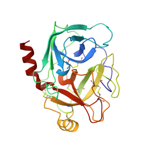
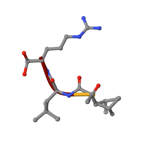
Two crystal structures of the leupeptin-trypsin complex.

Kurinov, I.V.Harrison, R.W.

(1996) Protein Sci 5: 752-758

  • DOI: https://doi.org/10.1002/pro.5560050420
  • Primary Citation of Related Structures:  
    1JRS, 1JRT

  • PubMed Abstract: 

    Three-dimensional structures of trypsin with the reversible inhibitor leupeptin have been determined in two different crystal forms. The first structure was determined at 1.7 A resolution with R-factor = 17.7% in the trigonal crystal space group P3(1)21, with unit cell dimensions of a = b = 55.62 A, c = 110.51 A. The second structure was determined at a resolution of 1.8 A with R-factor = 17.5% in the orthorhombic space group P2(1)2(1)2(1), with unit cell dimensions of a = 63.69 A, b = 69.37 A, c = 63.01 A. The overall protein structure is very similar in both crystal forms, with RMS difference for main-chain atoms of 0.27 A. The leupeptin backbone forms four hydrogen bonds with trypsin and a fifth hydrogen bond interaction is mediated by a water molecule. The aldehyde carbonyl of leupeptin forms a covalent bond of 1.42 A length with side-chain oxygen of Ser-195 in the active site. The reaction of trypsin with leupeptin proceeds through the formation of stable tetrahedral complex in which the hemiacetal oxygen atom is pointing out of the oxyanion hole and forming a hydrogen bond with His-57.


  • Organizational Affiliation

    Department of Pharmacology, Thomas Jefferson University, Philadelphia, Pennsylvania 19107, USA.


Macromolecules
Find similar proteins by:  (by identity cutoff)  |  3D Structure
Entity ID: 1
MoleculeChains Sequence LengthOrganismDetailsImage
TRYPSIN223Bos taurusMutation(s): 0 
EC: 3.4.21.4
UniProt
Find proteins for P00760 (Bos taurus)
Explore P00760 
Go to UniProtKB:  P00760
Entity Groups  
Sequence Clusters30% Identity50% Identity70% Identity90% Identity95% Identity100% Identity
UniProt GroupP00760
Sequence Annotations
Expand
  • Reference Sequence

Find similar proteins by:  Sequence   |   3D Structure  

Entity ID: 2
MoleculeChains Sequence LengthOrganismDetailsImage
Leupeptin4Streptomyces roseusMutation(s): 0 
EC: 3.4.21.4
Sequence Annotations
Expand
  • Reference Sequence
Small Molecules
Ligands 1 Unique
IDChains Name / Formula / InChI Key2D Diagram3D Interactions
CA
Query on CA

Download Ideal Coordinates CCD File 
C [auth A]CALCIUM ION
Ca
BHPQYMZQTOCNFJ-UHFFFAOYSA-N
Modified Residues  1 Unique
IDChains TypeFormula2D DiagramParent
AR7
Query on AR7
B
PEPTIDE-LIKEC6 H17 N4 O2ARG
Biologically Interesting Molecules (External Reference) 1 Unique
Entity ID: 2
IDChains NameType/Class2D Diagram3D Interactions
PRD_000216
Query on PRD_000216
B
LEUPEPTINOligopeptide / Enzyme inhibitor
Experimental Data & Validation

Experimental Data

  • Method: X-RAY DIFFRACTION
  • Resolution: 1.80 Å
  • R-Value Free: 0.207 
  • R-Value Work: 0.175 
  • R-Value Observed: 0.175 
  • Space Group: P 21 21 21
Unit Cell:
Length ( Å )Angle ( ˚ )
a = 63.686α = 90
b = 69.373β = 90
c = 63.013γ = 90
Software Package:
Software NamePurpose
PROCESSdata collection
X-PLORmodel building
X-PLORrefinement
PROCESSdata reduction
X-PLORphasing

Structure Validation

View Full Validation Report


Entry History 

Deposition Data

Revision History  (Full details and data files)

  • Version 1.0: 1996-10-14
    Type: Initial release
  • Version 1.1: 2008-03-24
    Changes: Version format compliance
  • Version 1.2: 2011-07-13
    Changes: Atomic model, Database references, Derived calculations, Non-polymer description, Structure summary, Version format compliance
  • Version 1.3: 2012-12-12
    Changes: Other