1JL4

CRYSTAL STRUCTURE OF THE HUMAN CD4 N-TERMINAL TWO DOMAIN FRAGMENT COMPLEXED TO A CLASS II MHC MOLECULE


Experimental Data Snapshot

  • Method: X-RAY DIFFRACTION
  • Resolution: 4.30 Å
  • R-Value Free: 0.453 
  • R-Value Work: 0.420 
  • R-Value Observed: 0.428 

wwPDB Validation   3D Report Full Report


This is version 1.4 of the entry. See complete history


Literature

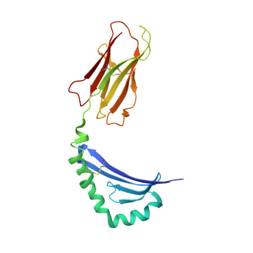
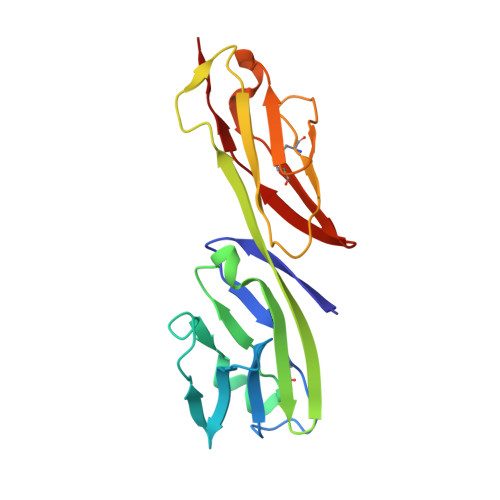
Crystal structure of the human CD4 N-terminal two-domain fragment complexed to a class II MHC molecule.

Wang, J.H.Meijers, R.Xiong, Y.Liu, J.H.Sakihama, T.Zhang, R.Joachimiak, A.Reinherz, E.L.

(2001) Proc Natl Acad Sci U S A 98: 10799-10804

  • DOI: https://doi.org/10.1073/pnas.191124098
  • Primary Citation of Related Structures:  
    1JL4

  • PubMed Abstract: 

    The structural basis of the interaction between the CD4 coreceptor and a class II major histocompatibility complex (MHC) is described. The crystal structure of a complex containing the human CD4 N-terminal two-domain fragment and the murine I-A(k) class II MHC molecule with associated peptide (pMHCII) shows that only the "top corner" of the CD4 molecule directly contacts pMHCII. The CD4 Phe-43 side chain extends into a hydrophobic concavity formed by MHC residues from both alpha 2 and beta 2 domains. A ternary model of the CD4-pMHCII-T-cell receptor (TCR) reveals that the complex appears V-shaped with the membrane-proximal pMHCII at the apex. This configuration excludes a direct TCR-CD4 interaction and suggests how TCR and CD4 signaling is coordinated around the antigenic pMHCII complex. Human CD4 binds to HIV gp120 in a manner strikingly similar to the way in which CD4 interacts with pMHCII. Additional contacts between gp120 and CD4 give the CD4-gp120 complex a greater affinity. Thus, ligation of the viral envelope glycoprotein to CD4 occludes the pMHCII-binding site on CD4, contributing to immunodeficiency.


  • Organizational Affiliation

    Laboratory of Immunobiology and Department of Cancer Immunology and AIDS, Dana-Farber Cancer Institute, Harvard Medical School, Boston, MA 02115, USA. jwang@dfci.harvard.edu


Macromolecules
Find similar proteins by:  (by identity cutoff)  |  3D Structure
Entity ID: 1
MoleculeChains Sequence LengthOrganismDetailsImage
H-2 CLASS II HISTOCOMPATIBILITY ANTIGEN, A-K ALPHA CHAIN178Mus musculusMutation(s): 0 
UniProt
Find proteins for P01910 (Mus musculus)
Explore P01910 
Go to UniProtKB:  P01910
Entity Groups  
Sequence Clusters30% Identity50% Identity70% Identity90% Identity95% Identity100% Identity
UniProt GroupP01910
Sequence Annotations
Expand
  • Reference Sequence
Find similar proteins by:  (by identity cutoff)  |  3D Structure
Entity ID: 2
MoleculeChains Sequence LengthOrganismDetailsImage
H-2 CLASS II HISTOCOMPATIBILITY ANTIGEN, A-K BETA CHAIN185Mus musculusMutation(s): 0 
UniProt
Find proteins for P06343 (Mus musculus)
Explore P06343 
Go to UniProtKB:  P06343
Entity Groups  
Sequence Clusters30% Identity50% Identity70% Identity90% Identity95% Identity100% Identity
UniProt GroupP06343
Sequence Annotations
Expand
  • Reference Sequence

Find similar proteins by:  Sequence   |   3D Structure  

Entity ID: 3
MoleculeChains Sequence LengthOrganismDetailsImage
OVOTRANSFERRIN16Gallus gallusMutation(s): 0 
UniProt
Find proteins for P02789 (Gallus gallus)
Explore P02789 
Go to UniProtKB:  P02789
Entity Groups  
Sequence Clusters30% Identity50% Identity70% Identity90% Identity95% Identity100% Identity
UniProt GroupP02789
Sequence Annotations
Expand
  • Reference Sequence
Find similar proteins by:  (by identity cutoff)  |  3D Structure
Entity ID: 4
MoleculeChains Sequence LengthOrganismDetailsImage
T-CELL SURFACE GLYCOPROTEIN CD4178Homo sapiensMutation(s): 0 
UniProt & NIH Common Fund Data Resources
Find proteins for P01730 (Homo sapiens)
Explore P01730 
Go to UniProtKB:  P01730
PHAROS:  P01730
GTEx:  ENSG00000010610 
Entity Groups  
Sequence Clusters30% Identity50% Identity70% Identity90% Identity95% Identity100% Identity
UniProt GroupP01730
Sequence Annotations
Expand
  • Reference Sequence
Experimental Data & Validation

Experimental Data

  • Method: X-RAY DIFFRACTION
  • Resolution: 4.30 Å
  • R-Value Free: 0.453 
  • R-Value Work: 0.420 
  • R-Value Observed: 0.428 
  • Space Group: P 43 2 2
Unit Cell:
Length ( Å )Angle ( ˚ )
a = 145.195α = 90
b = 145.195β = 90
c = 103.846γ = 90
Software Package:
Software NamePurpose
DENZOdata reduction
SCALEPACKdata scaling
AMoREphasing
EPMRphasing
FFFEARmodel building
REFMACrefinement
FFFEARphasing

Structure Validation

View Full Validation Report


Entry History 

Deposition Data

Revision History  (Full details and data files)

  • Version 1.0: 2001-09-19
    Type: Initial release
  • Version 1.1: 2008-04-27
    Changes: Version format compliance
  • Version 1.2: 2011-07-13
    Changes: Version format compliance
  • Version 1.3: 2017-10-04
    Changes: Refinement description
  • Version 1.4: 2023-08-16
    Changes: Data collection, Database references, Refinement description