1JKS

1.5A X-RAY STRUCTURE OF APO FORM OF A CATALYTIC DOMAIN OF DEATH-ASSOCIATED PROTEIN KINASE


Experimental Data Snapshot

  • Method: X-RAY DIFFRACTION
  • Resolution: 1.50 Å
  • R-Value Free: 0.202 
  • R-Value Work: 0.183 
  • R-Value Observed: 0.183 

wwPDB Validation   3D Report Full Report


This is version 1.3 of the entry. See complete history


Literature

Crystal structures of the catalytic domain of human protein kinase associated with apoptosis and tumor suppression.

Tereshko, V.Teplova, M.Brunzelle, J.Watterson, D.M.Egli, M.

(2001) Nat Struct Biol 8: 899-907

  • DOI: https://doi.org/10.1038/nsb1001-899
  • Primary Citation of Related Structures:  
    1IG1, 1JKK, 1JKL, 1JKS, 1JKT

  • PubMed Abstract: 

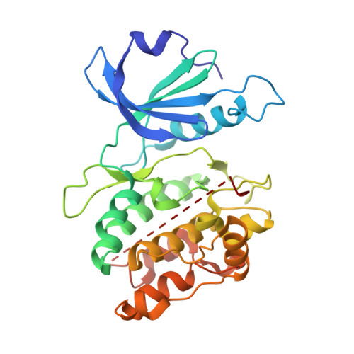
    We have determined X-ray crystal structures with up to 1.5 A resolution of the catalytic domain of death-associated protein kinase (DAPK), the first described member of a novel family of pro-apoptotic and tumor-suppressive serine/threonine kinases. The geometry of the active site was studied in the apo form, in a complex with nonhydrolyzable AMPPnP and in a ternary complex consisting of kinase, AMPPnP and either Mg2+ or Mn2+. The structures revealed a previously undescribed water-mediated stabilization of the interaction between the lysine that is conserved in protein kinases and the beta- and gamma-phosphates of ATP, as well as conformational changes at the active site upon ion binding. Comparison between these structures and nucleotide triphosphate complexes of several other kinases disclosed a number of unique features of the DAPK catalytic domain, among which is a highly ordered basic loop in the N-terminal domain that may participate in enzyme regulation.


  • Organizational Affiliation

    Department of Biological Sciences, Vanderbilt University, Nashville, Tennessee 37235, USA.


Macromolecules
Find similar proteins by:  (by identity cutoff)  |  3D Structure
Entity ID: 1
MoleculeChains Sequence LengthOrganismDetailsImage
DEATH-ASSOCIATED PROTEIN KINASE294Homo sapiensMutation(s): 0 
EC: 2.7.1
UniProt & NIH Common Fund Data Resources
Find proteins for P53355 (Homo sapiens)
Explore P53355 
Go to UniProtKB:  P53355
PHAROS:  P53355
GTEx:  ENSG00000196730 
Entity Groups  
Sequence Clusters30% Identity50% Identity70% Identity90% Identity95% Identity100% Identity
UniProt GroupP53355
Sequence Annotations
Expand
  • Reference Sequence
Experimental Data & Validation

Experimental Data

  • Method: X-RAY DIFFRACTION
  • Resolution: 1.50 Å
  • R-Value Free: 0.202 
  • R-Value Work: 0.183 
  • R-Value Observed: 0.183 
  • Space Group: P 21 21 21
Unit Cell:
Length ( Å )Angle ( ˚ )
a = 46.93α = 90
b = 62.55β = 90
c = 88.56γ = 90
Software Package:
Software NamePurpose
CNSrefinement
DENZOdata reduction
SCALEPACKdata scaling
CNSphasing

Structure Validation

View Full Validation Report


Entry History 

Deposition Data

Revision History  (Full details and data files)

  • Version 1.0: 2002-04-01
    Type: Initial release
  • Version 1.1: 2008-04-27
    Changes: Version format compliance
  • Version 1.2: 2011-07-13
    Changes: Version format compliance
  • Version 1.3: 2024-02-07
    Changes: Data collection, Database references