1JHT

Crystal structure of HLA-A2*0201 in complex with a nonameric altered peptide ligand (ALGIGILTV) from the MART-1/Melan-A.


Experimental Data Snapshot

  • Method: X-RAY DIFFRACTION
  • Resolution: 2.15 Å
  • R-Value Free: 0.252 
  • R-Value Work: 0.210 
  • R-Value Observed: 0.210 

wwPDB Validation   3D Report Full Report


This is version 1.3 of the entry. See complete history


Literature

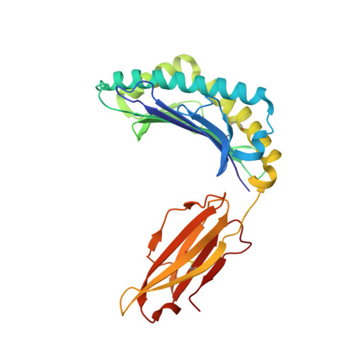
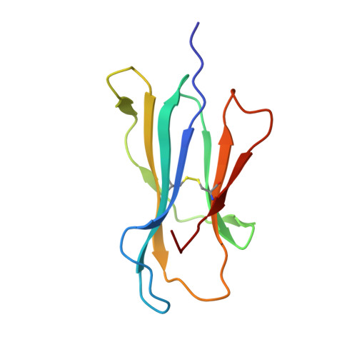
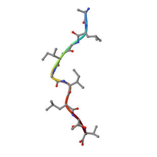
Crystal structures of two closely related but antigenically distinct HLA-A2/melanocyte-melanoma tumor-antigen peptide complexes.

Sliz, P.Michielin, O.Cerottini, J.C.Luescher, I.Romero, P.Karplus, M.Wiley, D.C.

(2001) J Immunol 167: 3276-3284

  • DOI: https://doi.org/10.4049/jimmunol.167.6.3276
  • Primary Citation of Related Structures:  
    1JF1, 1JHT

  • PubMed Abstract: 

    We have determined high-resolution crystal structures of the complexes of HLA-A2 molecules with two modified immunodominant peptides from the melanoma tumor-associated protein Melan-A/Melanoma Ag recognized by T cells-1. The two peptides, a decamer and nonamer with overlapping sequences (ELAGIGILTV and ALGIGILTV), are modified in the second residue to increase their affinity for HLA-A2. The modified decamer is more immunogenic than the natural peptide and a candidate for peptide-based melanoma immunotherapy. The crystal structures at 1.8 and 2.15 A resolution define the differences in binding modes of the modified peptides, including different clusters of water molecules that appear to stabilize the peptide-HLA interaction. The structures suggest both how the wild-type peptides would bind and how three categories of cytotoxic T lymphocytes with differing fine specificity might recognize the two peptides.


  • Organizational Affiliation

    Department of Molecular and Cellular Biology and Howard Hughes Medical Institute, Harvard University, Cambridge, MA 02138, USA.


Macromolecules
Find similar proteins by:  (by identity cutoff)  |  3D Structure
Entity ID: 1
MoleculeChains Sequence LengthOrganismDetailsImage
HLA CLASS I HISTOCOMPATIBILITY ANTIGEN, A-2 ALPHA CHAIN275Homo sapiensMutation(s): 0 
UniProt & NIH Common Fund Data Resources
Find proteins for P04439 (Homo sapiens)
Explore P04439 
Go to UniProtKB:  P04439
PHAROS:  P04439
GTEx:  ENSG00000206503 
Entity Groups  
Sequence Clusters30% Identity50% Identity70% Identity90% Identity95% Identity100% Identity
UniProt GroupP04439
Sequence Annotations
Expand
  • Reference Sequence
Find similar proteins by:  (by identity cutoff)  |  3D Structure
Entity ID: 2
MoleculeChains Sequence LengthOrganismDetailsImage
beta-2-microglobulin100Homo sapiensMutation(s): 0 
UniProt & NIH Common Fund Data Resources
Find proteins for P61769 (Homo sapiens)
Explore P61769 
Go to UniProtKB:  P61769
PHAROS:  P61769
GTEx:  ENSG00000166710 
Entity Groups  
Sequence Clusters30% Identity50% Identity70% Identity90% Identity95% Identity100% Identity
UniProt GroupP61769
Sequence Annotations
Expand
  • Reference Sequence

Find similar proteins by:  Sequence   |   3D Structure  

Entity ID: 3
MoleculeChains Sequence LengthOrganismDetailsImage
nonameric peptide ligand from the MART-1/Melan-A9N/AMutation(s): 0 
Sequence Annotations
Expand
  • Reference Sequence
Experimental Data & Validation

Experimental Data

  • Method: X-RAY DIFFRACTION
  • Resolution: 2.15 Å
  • R-Value Free: 0.252 
  • R-Value Work: 0.210 
  • R-Value Observed: 0.210 
  • Space Group: P 21 21 21
Unit Cell:
Length ( Å )Angle ( ˚ )
a = 49.19α = 90
b = 74.47β = 90
c = 122.29γ = 90
Software Package:
Software NamePurpose
SCALEPACKdata scaling
MOLREPphasing
CNSrefinement

Structure Validation

View Full Validation Report


Entry History 

Deposition Data

Revision History  (Full details and data files)

  • Version 1.0: 2001-09-14
    Type: Initial release
  • Version 1.1: 2008-04-27
    Changes: Version format compliance
  • Version 1.2: 2011-07-13
    Changes: Version format compliance
  • Version 1.3: 2023-08-16
    Changes: Data collection, Database references, Refinement description