1JGR

Crystal Structure Analysis of the B-DNA Dodecamer CGCGAATTCGCG with Thallium Ions.


Experimental Data Snapshot

  • Method: X-RAY DIFFRACTION
  • Resolution: 1.20 Å
  • R-Value Free: 0.222 
  • R-Value Work: 0.169 
  • R-Value Observed: 0.163 

wwPDB Validation   3D Report Full Report


This is version 1.3 of the entry. See complete history


Literature

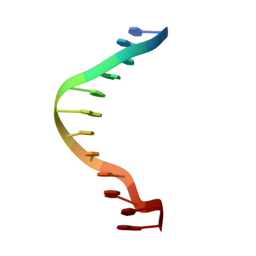
Locating monovalent cations in the grooves of B-DNA.

Howerton, S.B.Sines, C.C.VanDerveer, D.Williams, L.D.

(2001) Biochemistry 40: 10023-10031

  • DOI: https://doi.org/10.1021/bi010391+
  • Primary Citation of Related Structures:  
    1JGR

  • PubMed Abstract: 

    Here we demonstrate that monovalent cations can localize around B-DNA in geometrically regular, sequence-specific sites in oligonucleotide crystals. Positions of monovalent ions were determined from high-resolution X-ray diffraction of DNA crystals grown in the presence of thallium(I) cations (Tl(+)). Tl(+) has previously been shown to be a useful K(+) mimic. Tl(+) positions determined by refinement of model to data are consistent with positions determined using isomorphous F(Tl) - F(K) difference Fouriers and anomalous difference Fouriers. None of the observed Tl(+) sites surrounding CGCGAATTCGCG are fully occupied by Tl(+) ions. The most highly occupied sites, located within the G-tract major groove, have estimated occupancies ranging from 20% to 35%. The occupancies of the minor groove sites are estimated to be around 10%. The Tl(+) positions in general are not in direct proximity to phosphate groups. The A-tract major groove appears devoid of localized cations. The majority of the observed Tl(+) ions interact with a single duplex and so are not engaged in lattice interactions or crystal packing. The locations of the cation sites are dictated by coordination geometry, electronegative potential, avoidance of electropositive amino groups, and cation-pi interactions. It appears that partially dehydrated monovalent cations, hydrated divalent cations, and polyamines compete for a common binding region on the floor of the G-tract major groove.


  • Organizational Affiliation

    School of Chemistry and Biochemistry, Georgia Institute of Technology, Atlanta, Georgia 30332-0400, USA.


Macromolecules

Find similar nucleic acids by:  Sequence   |   3D Structure  

Entity ID: 1
MoleculeChains LengthOrganismImage
5'-D(*CP*GP*CP*GP*AP*AP*TP*TP*CP*GP*CP*G)-3'
A, B
12N/A
Sequence Annotations
Expand
  • Reference Sequence
Small Molecules
Ligands 2 Unique
IDChains Name / Formula / InChI Key2D Diagram3D Interactions
TL
Query on TL

Download Ideal Coordinates CCD File 
D [auth A]
E [auth A]
F [auth A]
G [auth A]
H [auth A]
D [auth A],
E [auth A],
F [auth A],
G [auth A],
H [auth A],
I [auth A],
J [auth A],
K [auth B],
L [auth B],
M [auth B],
N [auth B],
O [auth B],
P [auth B]
THALLIUM (I) ION
Tl
ZLUSCZLCHQSJRU-UHFFFAOYSA-N
MG
Query on MG

Download Ideal Coordinates CCD File 
C [auth A]MAGNESIUM ION
Mg
JLVVSXFLKOJNIY-UHFFFAOYSA-N
Experimental Data & Validation

Experimental Data

  • Method: X-RAY DIFFRACTION
  • Resolution: 1.20 Å
  • R-Value Free: 0.222 
  • R-Value Work: 0.169 
  • R-Value Observed: 0.163 
  • Space Group: P 21 21 21
Unit Cell:
Length ( Å )Angle ( ˚ )
a = 25.944α = 90
b = 40.744β = 90
c = 66.202γ = 90
Software Package:
Software NamePurpose
SHELXL-97refinement
MOSFLMdata reduction
CCP4data scaling

Structure Validation

View Full Validation Report


Entry History 

Deposition Data

Revision History  (Full details and data files)

  • Version 1.0: 2001-09-05
    Type: Initial release
  • Version 1.1: 2008-04-27
    Changes: Version format compliance
  • Version 1.2: 2011-07-13
    Changes: Version format compliance
  • Version 1.3: 2023-08-16
    Changes: Data collection, Database references, Derived calculations, Refinement description