1JBG

Crystal Structure of MtaN, the Bacillus subtilis Multidrug Transporter Activator, N-terminus


Experimental Data Snapshot

  • Method: X-RAY DIFFRACTION
  • Resolution: 2.75 Å
  • R-Value Free: 0.287 
  • R-Value Work: 0.228 
  • R-Value Observed: 0.231 

wwPDB Validation   3D Report Full Report


This is version 1.3 of the entry. See complete history


Literature

Crystal structure of MtaN, a global multidrug transporter gene activator.

Godsey, M.H.Baranova, N.N.Neyfakh, A.A.Brennan, R.G.

(2001) J Biol Chem 276: 47178-47184

  • DOI: https://doi.org/10.1074/jbc.M105819200
  • Primary Citation of Related Structures:  
    1JBG

  • PubMed Abstract: 

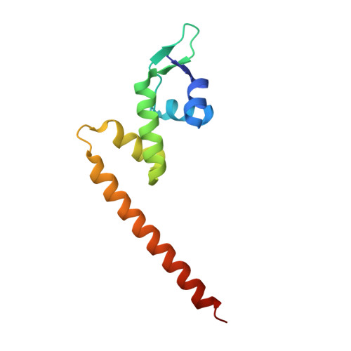
    MtaN (Multidrug Transporter Activation, N terminus) is a constitutive, transcriptionally active 109-residue truncation mutant, which contains only the N-terminal DNA-binding and dimerization domains of MerR family member Mta. The 2.75 A resolution crystal structure of apo-MtaN reveals a winged helix-turn-helix protein with a protruding 8-turn helix (alpha5) that is involved in dimerization by the formation of an antiparallel coiled-coil. The hydrophobic core and helices alpha1 through alpha4 are structurally homologous to MerR family member BmrR bound to DNA, whereas one wing (Wing 1) is shifted. Differences between the orientation of alpha5 with respect to the core and the revolution of the antiparallel coiled-coil lead to significantly altered conformations of MtaN and BmrR dimers. These shifts result in a conformation of MtaN that appears to be incompatible with the transcription activation mechanism of BmrR and suggest that additional DNA-induced structural changes are necessary.


  • Organizational Affiliation

    Department of Biochemistry and Molecular Biology, Oregon Health & Science University, Portland, OR 97201-3098, USA.


Macromolecules
Find similar proteins by:  (by identity cutoff)  |  3D Structure
Entity ID: 1
MoleculeChains Sequence LengthOrganismDetailsImage
transcription activator of multidrug-efflux transporter genes mta109Bacillus subtilisMutation(s): 0 
UniProt
Find proteins for P71039 (Bacillus subtilis (strain 168))
Explore P71039 
Go to UniProtKB:  P71039
Entity Groups  
Sequence Clusters30% Identity50% Identity70% Identity90% Identity95% Identity100% Identity
UniProt GroupP71039
Sequence Annotations
Expand
  • Reference Sequence
Experimental Data & Validation

Experimental Data

  • Method: X-RAY DIFFRACTION
  • Resolution: 2.75 Å
  • R-Value Free: 0.287 
  • R-Value Work: 0.228 
  • R-Value Observed: 0.231 
  • Space Group: I 21 21 21
Unit Cell:
Length ( Å )Angle ( ˚ )
a = 49.382α = 90
b = 67.838β = 90
c = 115.005γ = 90
Software Package:
Software NamePurpose
CNSrefinement
MOSFLMdata reduction
CCP4data scaling
CNSphasing

Structure Validation

View Full Validation Report


Entry History 

Deposition Data

Revision History  (Full details and data files)

  • Version 1.0: 2001-11-28
    Type: Initial release
  • Version 1.1: 2008-04-27
    Changes: Version format compliance
  • Version 1.2: 2011-07-13
    Changes: Derived calculations, Version format compliance
  • Version 1.3: 2024-02-07
    Changes: Data collection, Database references